


3.) Protein Story Phase: Insulin
Climbing the Mountain
Insulin is a protein hormone involved in regulating the concentration of sugar in our bloodstream.
The resources below will introduce the basics of insulin structure and function. You will then choose a specific protein story related to insulin that you want to model. You will read a scientific research article, find an insulin protein structure file, and design a 3D printed physical model of an insulin protein.
Note: If you skipped the Training Phase and need to review basic protein structure-function concepts, visit the Training Phase Protein Structure Resources.
Recommended Models for the Insulin Protein Story Phase
The MAPS program revolves around using physical models as teaching tools. So if at all possible, we encourage you to use the recommeneded models below for the insulin protein story phase.
All recommended models are available to borrow free of charge through the MSOE Lending Library, or are available for purchase through 3D Molecular Designs. Use the discount code below to receive a 10% MAPS discount on your 3D Molecular Designs purchases.

1. Introduction to Insulin
In this section, we will explore the basic structure and functions of insulins, including why we need insulin and how it regulates sugar in the blood.
An Introduction to Insulin2. Insulin Structure
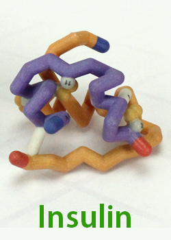
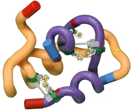
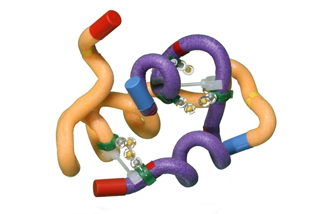
In this section we are going to explore the structure of insulin. This key hormone is a two chained protein with both alpha helical regions and a strand of beta pleated sheet.
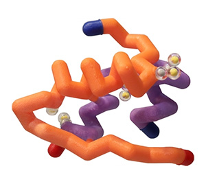
Modeling Insulin StructureWe are going to start out by modeling the basic structure of an insulin molecule. As you will see, it has a very unique structure. In order to do this, you will need access to the 3DMD mRNA to Protein Insulin Folding Kit (available for purchase or loan), some toobers and cysteine sidechains, or other materials that you might think of (pipe cleaners, etc).
If you do not have access to the kit, you will need toobers or other materials to model the two peptide backbones: A chain = 21 in; B chain = 30 in. You will need to add cysteine sidechains, alpha helices and beta sheets according to the map below.
Now you need to fold the 2 chains in the proper structure. The A chain will look like an upside down "U" with a flat section between the 2 helices. The B chain looks like a "Z" with the alpha helix being the diagonal section. The A chain will have one intrachain disulfide bond between A6 and A11. Then there will be 2 interchain disulfide bonds; one between A6 and B6 and one between A20 and B19.
Use the handout below to help you explore and model insulin. Take your time, ask lots of questions, discuss, and have fun. I'll summarize later in a video.
Thought question: What "level" of protein structure did you just model? Primary, secondary, tertiary, quaternary? Discuss!

Insulin Folding Guidelines
Modeling Insulin Transcription and Translation
So now that you have a good idea of the structure of insulin, you might be asking yourself a lot of questions. For example, why 2 chains? Why so many disulfide bonds? How do the the correct cysteines find each other?
First, make sure that your team understanding the "Central Dogma of Molecular Biology". That's just a stuffy way of saying that DNA is the code for messenger RNA (or mRNA) which is the code for a protein. If your teacher has the 3DMD Flow if Genetic information Kit, you can review these processes with that. Also, take a look at the RNA coding activity handout below. You can use either the attached codon chart or circle (your choice) to "transcribe" DNA to RNA and "translate" RNA to protein. In other words, you will "crack the code" of the insulin gene!
mRNA Coding ActivityGenetic Codon Chart
Mini Circle Codon Chart
Modeling Insulin Synthesis and Processing
Now that you understand how the DNA within the insulin gene encodes the RNA and the RNA encodes the sequence for the insulin protein, it is time to model how insulin is synthesized and processed. The mRNA map below show the fully transcribed mRNA and the 3 possible reading frames for protein translation. As you go through the handout below using the mRNA map as a reference, you might want to model insulin processing with different colored pipe cleaners twisted together.
But first, let's make sure you familiar with the Golgi so that you can understand its role in insulin processing. I'm going to steal one of Science with Tom's lyrics for MAPS:
"The cell does a lot, so we're gonna model it, model it, model model like it's hot!

Insulin Synthesis Handout
Insulin Storage and Secretion
After modeling insulin synthesis and processing, you should now understand that the insulin translation product is called preproinsulin. This includes the A and the B chain that we modeled earlier and 2 peptide segments that are not found in the final product, the signal peptide and the C peptide. This precursor protein needs to be processed into the final product which is then secreted by the pancreatic beta cells.
The modeling activity you just did gave you a simplistic idea of how that might happen. Turns out it is a little more complicated than that. I found this figure in the review I've attached below. You can look at the review if you are interested but I thought the figure was cool. Take a close look at this figure and make some observations. Does anything surprise you about this figure? Without reading the paper, can you use the knowledge of insulin synthesis that you have constructed so far (along with anything you might remember from Biology class ;)) to make some statements that describe what is being depicted here? Don't worry about understanding all of it or being "correct". There is a lot of detail here. Just see what looks familiar and try to fit it in to what you think you know. Discuss as a team how you think the system might work based on this figure. We'll discuss in the upcoming summary video.
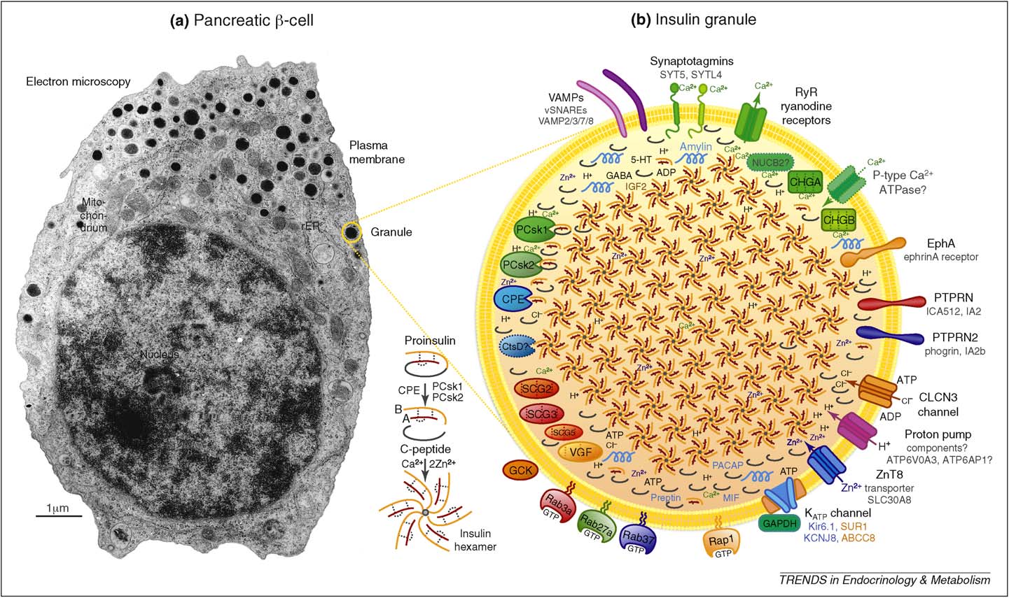
Insulin Secretion Signaling Paper
3. Insulin Action and Engineering
Glucose in the blood stimulates the pancreas to release insulin. Insulin acts on different tissues in different ways, depending upon the needs of the tissue. Muscle utilizes glucose for energy and liver and adipose tissues have the ability to store glucose for later use.
Insulin Release and Activation Pathways4. Defining Your Insulin Protein Story
Now that you are armed with an extensive understanding of what insulin does and how its structure is important to its function, it is time to decide what specific insulin story you would like to explore.
The expandable content below will give you some suggested topics you may choose to focus on, recommended insulin research papers and reviews to read, and link to a collection of insulin structure files to use when designing your 3D printed protein model.
Insulin Topics and Review Articles
Once you have defined the insulin protein story you want to tell, you will need to find a protein structure file that you can work with in Jmol to design your 3D printed insulin model.
Designing and building your own physical insulin model will give you great insight into specific structures that are important to the insulin protein story you have chosen to focus on. Even if you don't have your design 3D printed, studying the 3D design in specialized computer software is still helpful.
Below are a few additional links that may help you in your insulin structure search.
What Does it Mean to "Design" a Protein Model?
Tim Herman, PhD Explains How to Approach Your Model Design in Jmol
"Designing" a protein model means you explore a protein structure in Jmol, and then simplify the way the protein is visually displayed to make the key features of the protein that help communicate your molecular story more obvious.
This can mean hiding some atoms that are not important for your protein story, changing the display format of certain parts of your protein structure or changing colors to best highlight the most important parts of the protein structures. In the next section, you will learn the Jmol commands needed to accomplish this.
How to Approach Your Model Design
Tim Herman, PhD Explains How to Approach Your Model Design in Jmol
All 3D printed protein models made in the MAPS program are designed using the program Jmol. You start with a protein structure file (.PDB file), which contains the 3D locations of all of the atoms that make up the protein. Using the Jmol commands you will learn below, you can then edit the way the protein is displayed, hiding some atoms, changing display formats and customizing colors. When happy with your design, you can export your protein model from Jmol in file formats suitable for 3D printing.
We have created a detailed Jmol Training Guide that will cover everything you need to know to design and build your model. The Jmol Training Guide is broken down into four main sections, which we strongly recommend you explore in order until you are comfortable with Jmol and designing protein models for 3D printing.
- 1) Getting Started:
- Accessing Jmol
- Loading Structures
- Saving and Reloading - 2) Simplifying Your Protein:
- Exploring the Structure
- Focusing on the Backbone - 3) Adding the Details:
- Adding Sidechains
- Adding Ligands
- Adding Molecular Bonds - 4) 3D Printing Your Design:
- Adding Support Struts
- Checking Your Design
- Print it Yourself
- Pay Someone Else to Print it
Moving to the Next Phase
Once your team has finished the protein story phase, move on the Capstone Experience, where you will learn how to present your project using your 3D printed model and a variety of complimentary digital and print media.