


3.) Protein Story Phase: Aquaporin
Climbing the Mountain
Aquaporin is a fascinating protein involved in regulating the flow of water across a membrane.
The resources below will introduce membranes and the basics of aquaporin structure and function. You will then choose a specific protein story related to aquaporin that you want to model. You will read a scientific research article, find an aquaporin protein structure file, and design a 3D printed physical model of aquaporin.
Note: If you skipped the Training Phase and need to review basic protein structure-function concepts, visit the Training Phase Protein Structure Resources.
Recommended Models for the Aquaporin Protein Story Phase
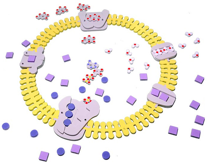
The MAPS program revolves around using physical models as teaching tools. So if at all possible, we encourage you to use the recommended models below for the aquaporin protein story phase.
All recommended models are available to borrow free of charge through the MSOE Lending Library, or are available for purchase through 3D Molecular Designs. Use the discount code below to receive a 10% MAPS discount on your 3D Molecular Designs purchases.

1. Introduction to Membranes
In order to understand the need for a water channel like aquaporin, we first need to introduce some fascinating molecules called phospholipids. Click below to view expandable content that will discuss phospholipids and plasma membranes.
Exploring Phospholipids and Plasma Membranes
Plasma Membranes form barriers in living things, separating the inside from the outside of cells, as well as separating different compartments, called organelles, inside of a single cell. Plasma membranes are comprised of individual phospholipids, which come together to form a phospholipid bilayer
This will be a lot more fun with the use of physical models! So if at all possible, we encourage you to use the Phospholipid and Membrane Transport Kit for this section.
- The Pospholipid and Membrane Transport Kit is available to borrow (free of charge) from the MSOE Lending Library.
- The Pospholipid and Membrane Transport Kit is available to purchase from the company 3D Molecular Designs.
Take your time with these activities! There is a lot to think about. Don’t be afraid to ask each other questions, as the more you question, the more you learn! The links below will provide you with additional activities to help you work with the Phospholipid and Membrane Transport Kit, and the video below will summarize the key attributes of phospholipids as they relate to aquaporin.
- Pospholipid and Membrane Transport Kit Brief
- Full Pospholipid Lesson
- Full Membrane Lesson
- Full Membrane Transport Lesson
- Additional Pospholipid and Membrane Transport Kit Resources
Summary of Phospholipids and Membranes
Summary video on the key points of phospholipids and membranes
2. The Structure and Function of Aquaporin Proteins
In this section we are going to learn how the protein structure that are unique to aquaporins allow only water molecules to pass through them.
Below is a collection of expandable content that will introduce the structure of aquaporins and how they function.
Peter Agre and the Discovery of Aquaporin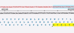
In this section we will explore the sequence of amino acids that make up the aquaporin protein, and how that relates to the overall shape and structure of aquaporin. Start by reading the research paper Structural determinants of water permeation through aquaporin-1 by Kazuyoshi Murata, Et al., and take some time to discuss the important features of the aquaporin-1 channel that the authors described.
Here is where the fun begins. Next you should print out and assemble the aqauporin gene map linked to below, and, after reading the Murata paper, begin to correlate aquaporin sequence to structure. There is no right way or wrong way. Just take your time and work together to try to make sense of the research paper and gene map as much as possible.
You can use the worksheet linked below as a guide, which will help you figure out how the aquaporin amino acid sequence is folded into secondary, tertiary, and quaternary structure, and how the structure is critical for aquaporin to be an effective water channel.
3. Defining Your Aquaporin Protein Story
Now that you are armed with an extensive understanding of what aquaporins are, how certain evolutionarily conserved sequences are important to folding them into their unique structure, and how their structure enables their critical function, it is time to decide what specific aquaporin story you would like to explore.
The expandable content below will give you some suggested topics you may choose to focus on, recommended aquaporin research papers and reviews to read, and link to a collection of aquaporin structure files to use when designing your 3D printed protein model.
Aquaporin Topics and Review Articles
Once you have defined the aquaporin protein story you want to tell, you will need to find a protein structure file that you can work with in Jmol to design your 3D printed aquaporin model.
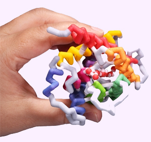
Designing and building your own physical aquaporin model will give you great insight into specific structures that are important to the aquaporin protein story you have chosen to focus on. Even if you don't have your design 3D printed, studying the 3D design in specialized computer software is still helpful.
Note that not all isoforms of aquaporin have public structure files, and while new structures are being discovered every day, below are a collection of recommended structures, along with their corresponding structure research paper
AQP4 Papers
- 3GD8 Structure Paper
- 3GD8 Supplement Paper
- 3IYZ Structure Paper
- Aggregation state determines the localization and function of M1– and M23–aquaporin-4 in astrocytes
- Super-resolution imaging of aquaporin-4 orthogonal arrays of particles in cell membranes
- Aquaporin-4 orthogonal arrays of particles from a physiological and pathophysiological point of view
AQP5 Papers
Below are a few additional links that may help you in your aquaporin structure search.
What Does it Mean to "Design" a Protein Model?
Tim Herman, PhD Explains How to Approach Your Model Design in Jmol
"Designing" a protein model means you explore a protein structure in Jmol, and then simplify the way the protein is visually displayed to make the key features of the protein that help communicate your molecular story more obvious.
This can mean hiding some atoms that are not important for your protein story, changing the display format of certain parts of your protein structure or changing colors to best highlight the most important parts of the protein structures. In the next section, you will learn the Jmol commands needed to accomplish this.
How to Approach Your Model Design
Tim Herman, PhD Explains How to Approach Your Model Design in Jmol
All 3D printed protein models made in the MAPS program are designed using the program Jmol. You start with a protein structure file (.PDB file), which contains the 3D locations of all of the atoms that make up the protein. Using the Jmol commands you will learn below, you can then edit the way the protein is displayed, hiding some atoms, changing display formats and customizing colors. When happy with your design, you can export your protein model from Jmol in file formats suitable for 3D printing.
We have created a detailed Jmol Training Guide that will cover everything you need to know to design and build your model. The Jmol Training Guide is broken down into four main sections, which we strongly recommend you explore in order until you are comfortable with Jmol and designing protein models for 3D printing.
- 1) Getting Started:
- Accessing Jmol
- Loading Structures
- Saving and Reloading - 2) Simplifying Your Protein:
- Exploring the Structure
- Focusing on the Backbone - 3) Adding the Details:
- Adding Sidechains
- Adding Ligands
- Adding Molecular Bonds - 4) 3D Printing Your Design:
- Adding Support Struts
- Checking Your Design
- Print it Yourself
- Pay Someone Else to Print it
Moving to the Next Phase
Once your team has finished the protein story phase, move on the Capstone Experience, where you will learn how to present your project using your 3D printed model and a variety of complimentary digital and print media.