Gallery of Physical Molecular Models
Phospholipids with Other Molecules

Phospholipids with Other Molecules
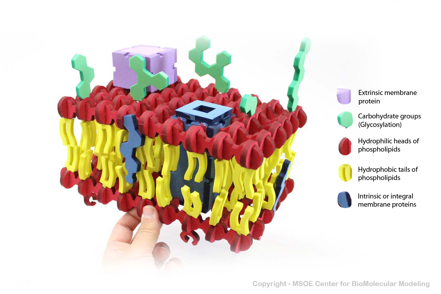
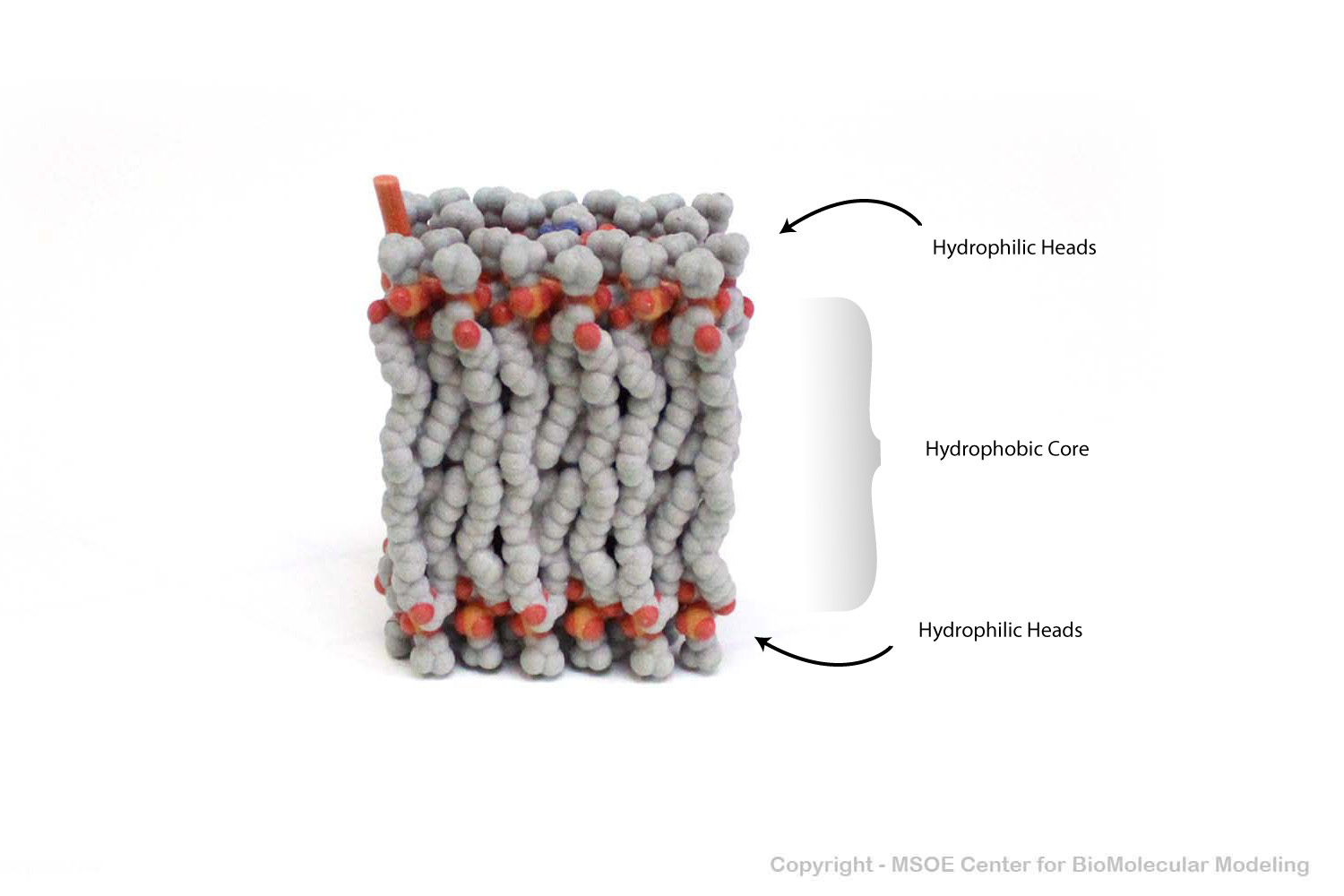
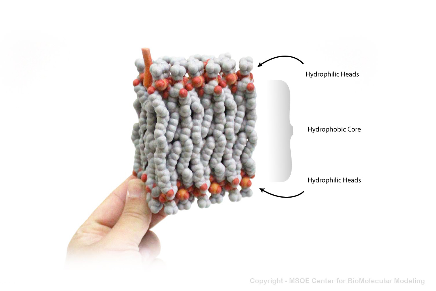
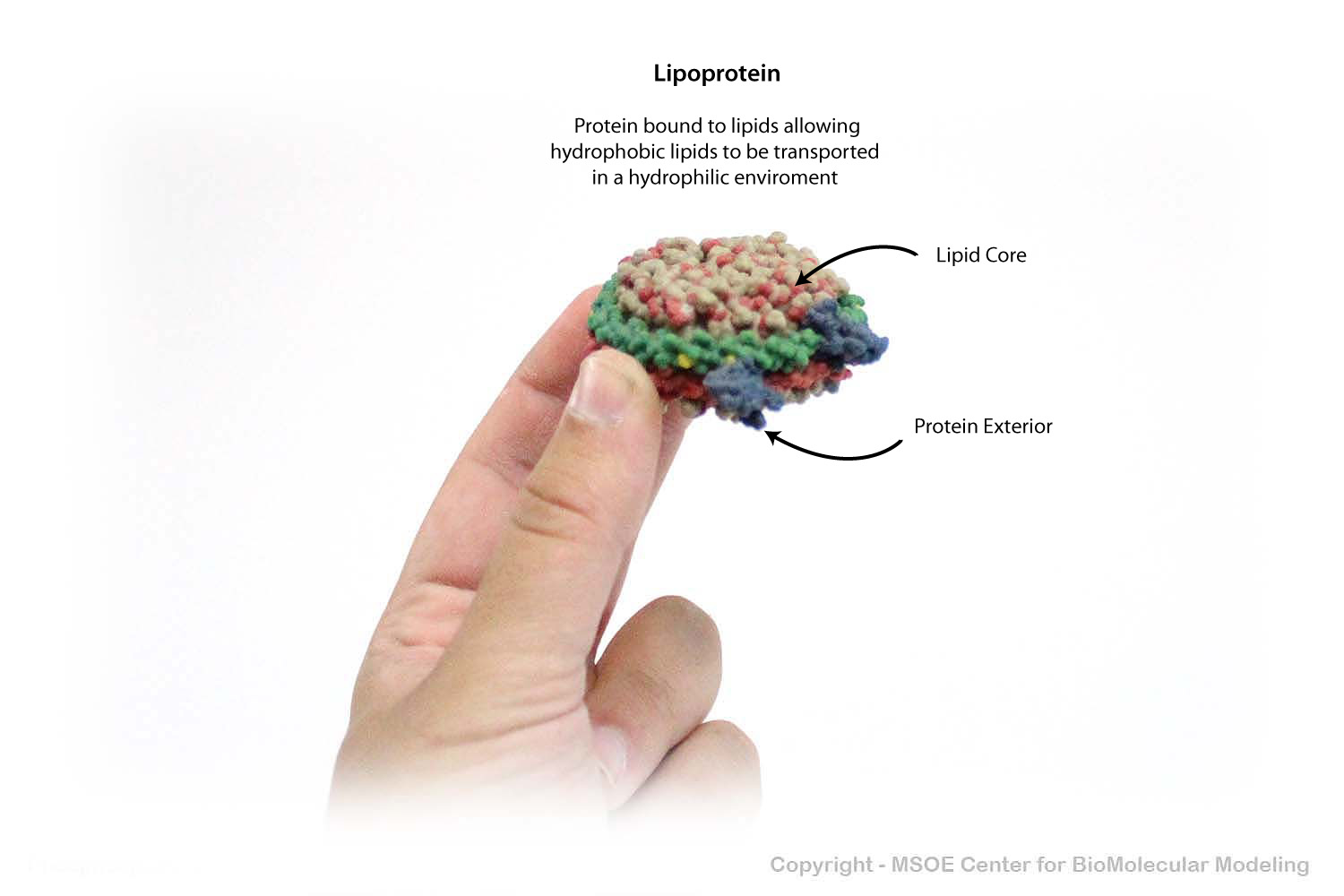
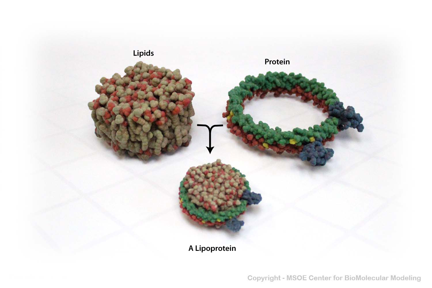
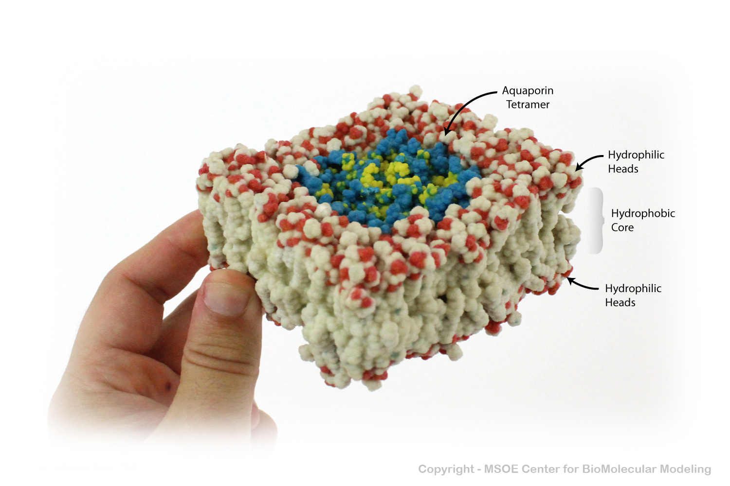
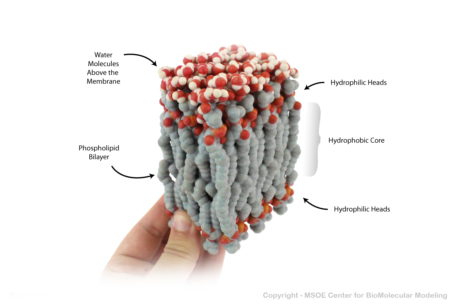
Multiple phospholipids can aggregate into a lipid bilayer, with hydrophobic tails in the middle of the bilayer and hydrophilic heads on the outside of the bilayer. Other molecules, such as protein and glycans decorate the bilayer.
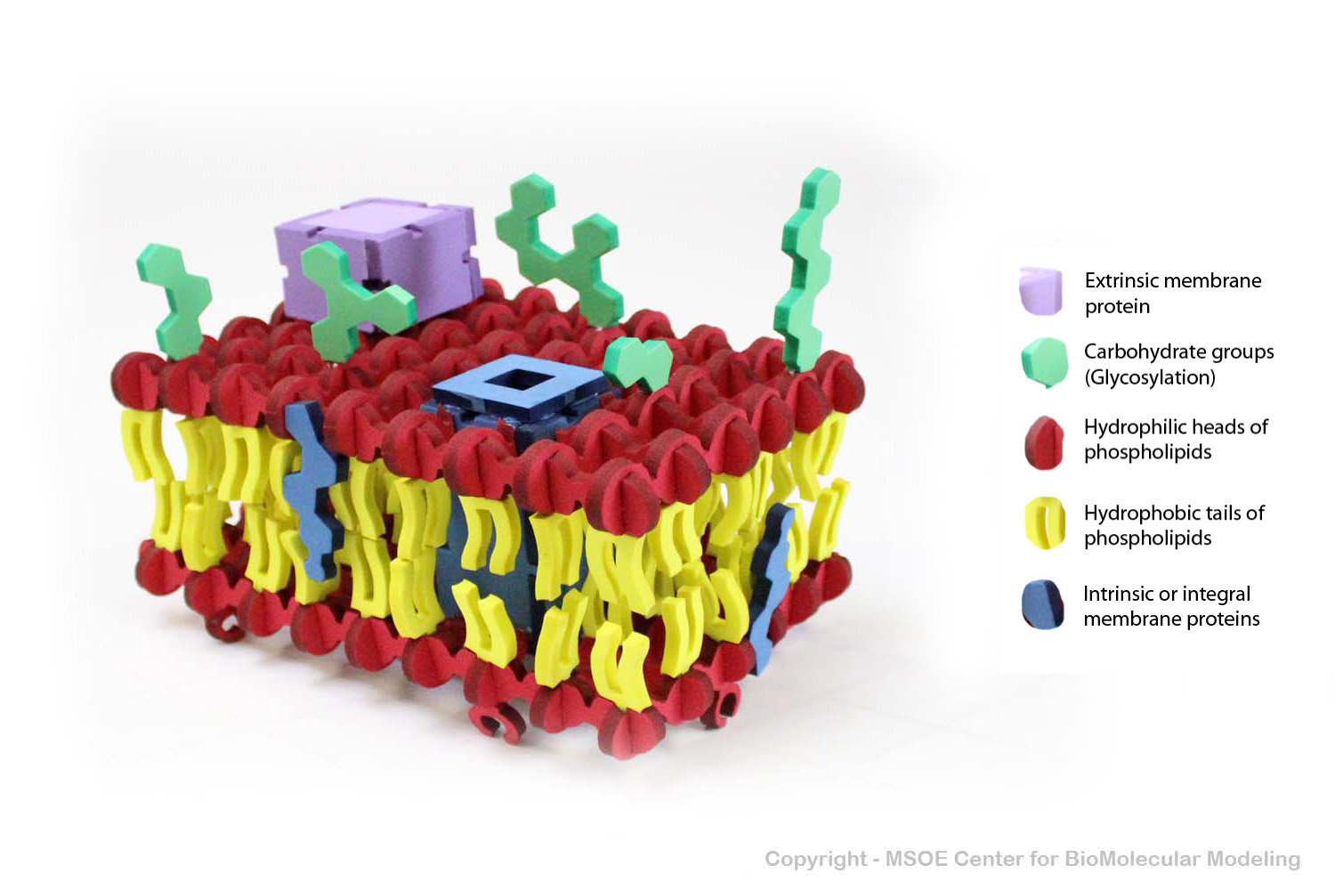
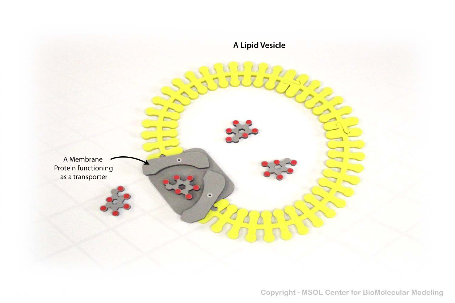
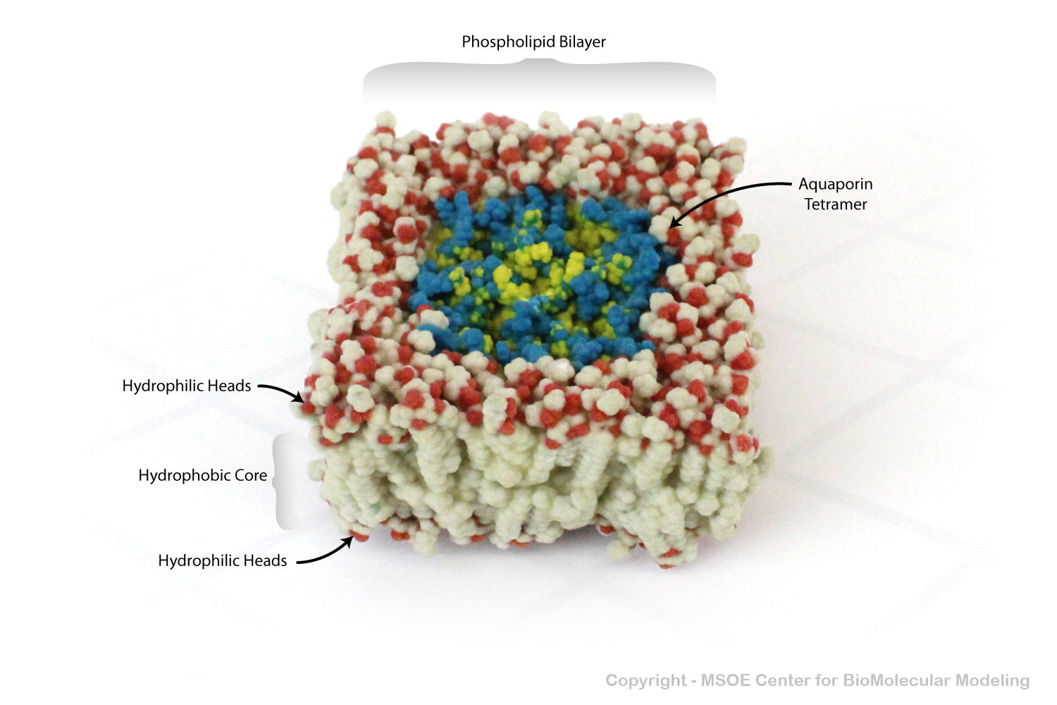
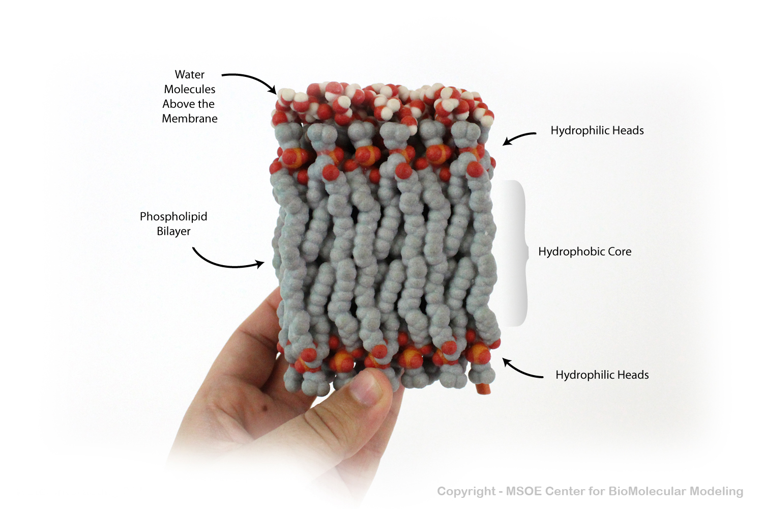
Multiple phospholipids can aggregate into a lipid bilayer, with hydrophobic tails in the middle of the bilayer and hydrophilic heads on the outside of the bilayer. Other molecules, such as protein and glycans decorate the bilayer.
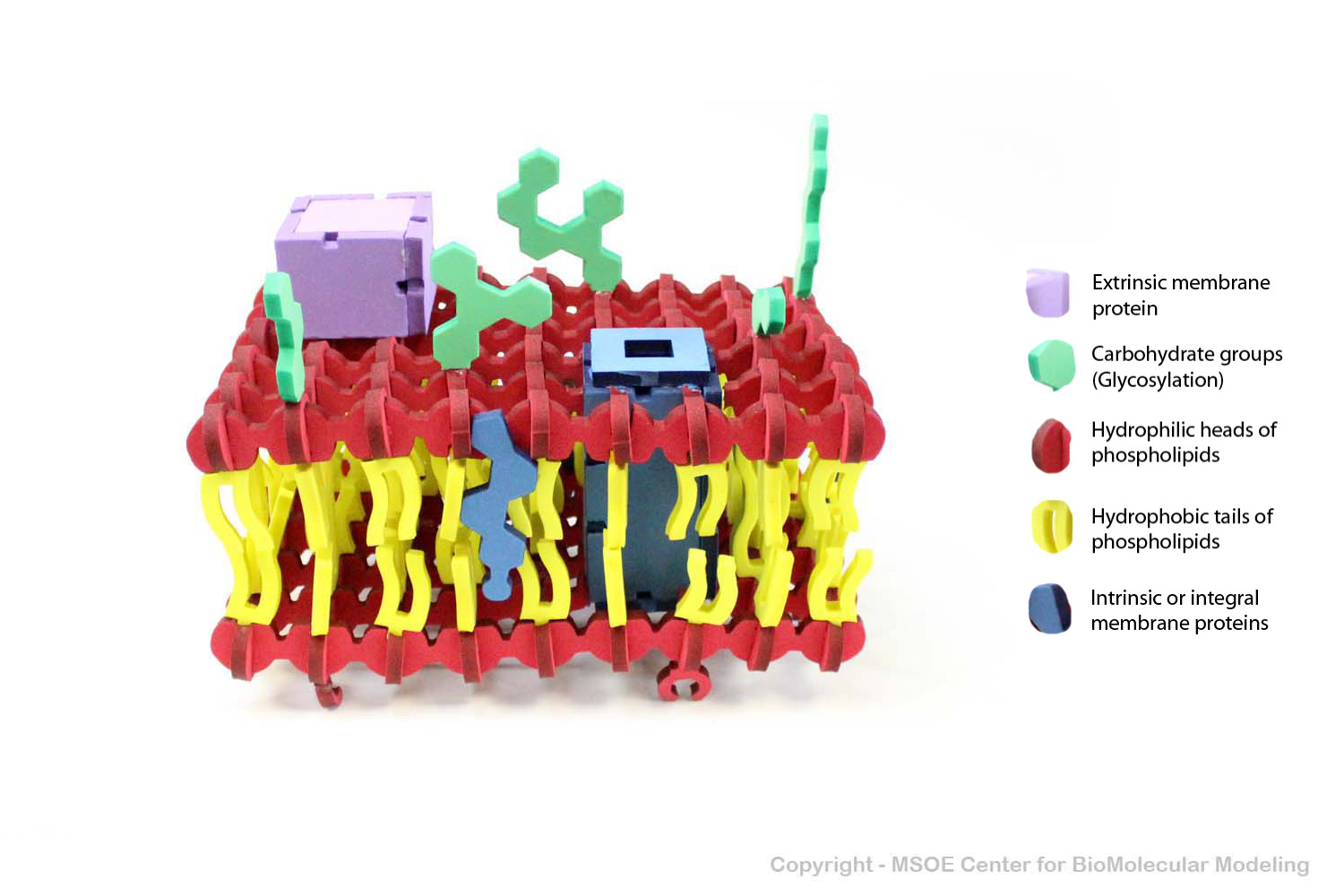
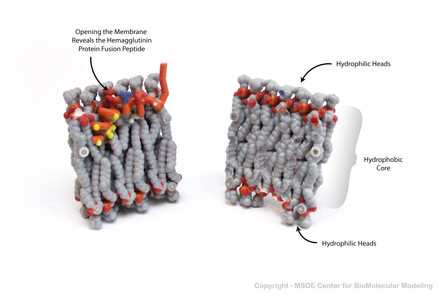
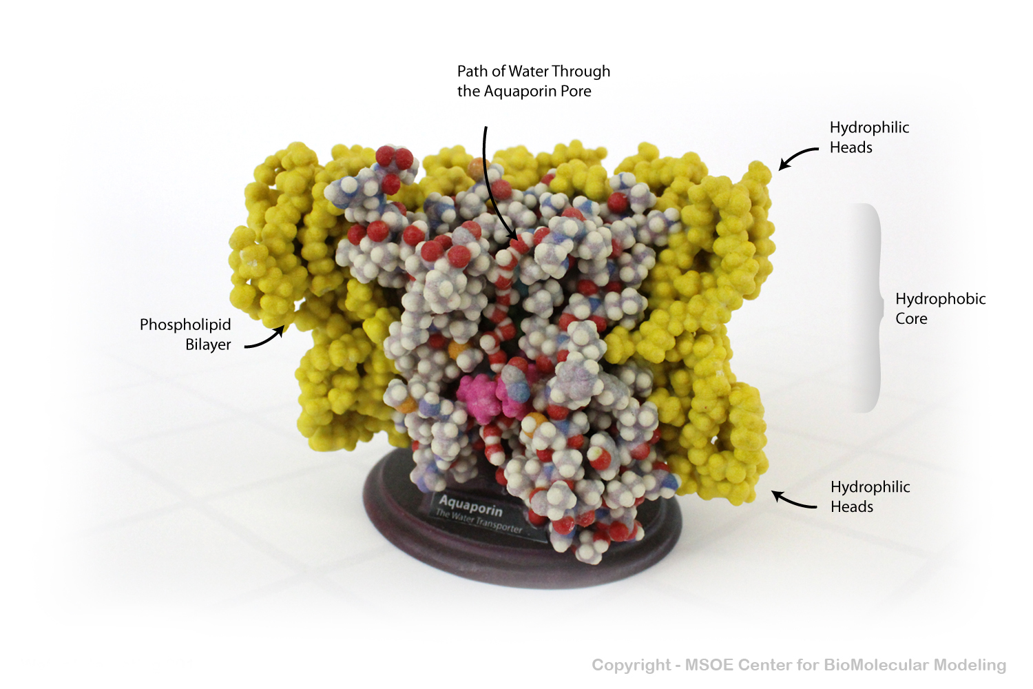
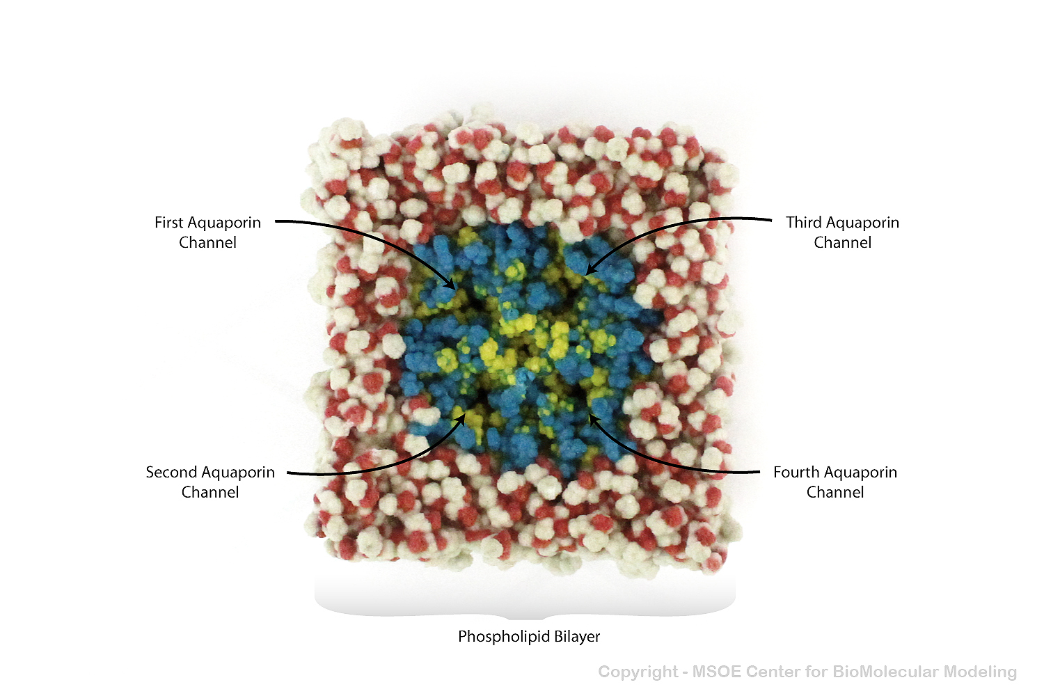
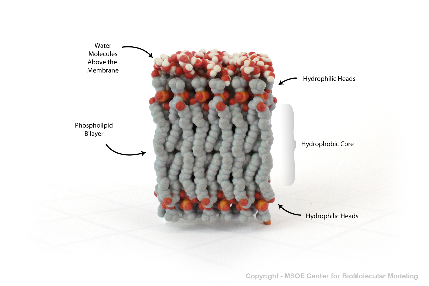
Multiple phospholipids can aggregate into a lipid bilayer, with hydrophobic tails in the middle of the bilayer and hydrophilic heads on the outside of the bilayer. Other molecules, such as protein and glycans decorate the bilayer.
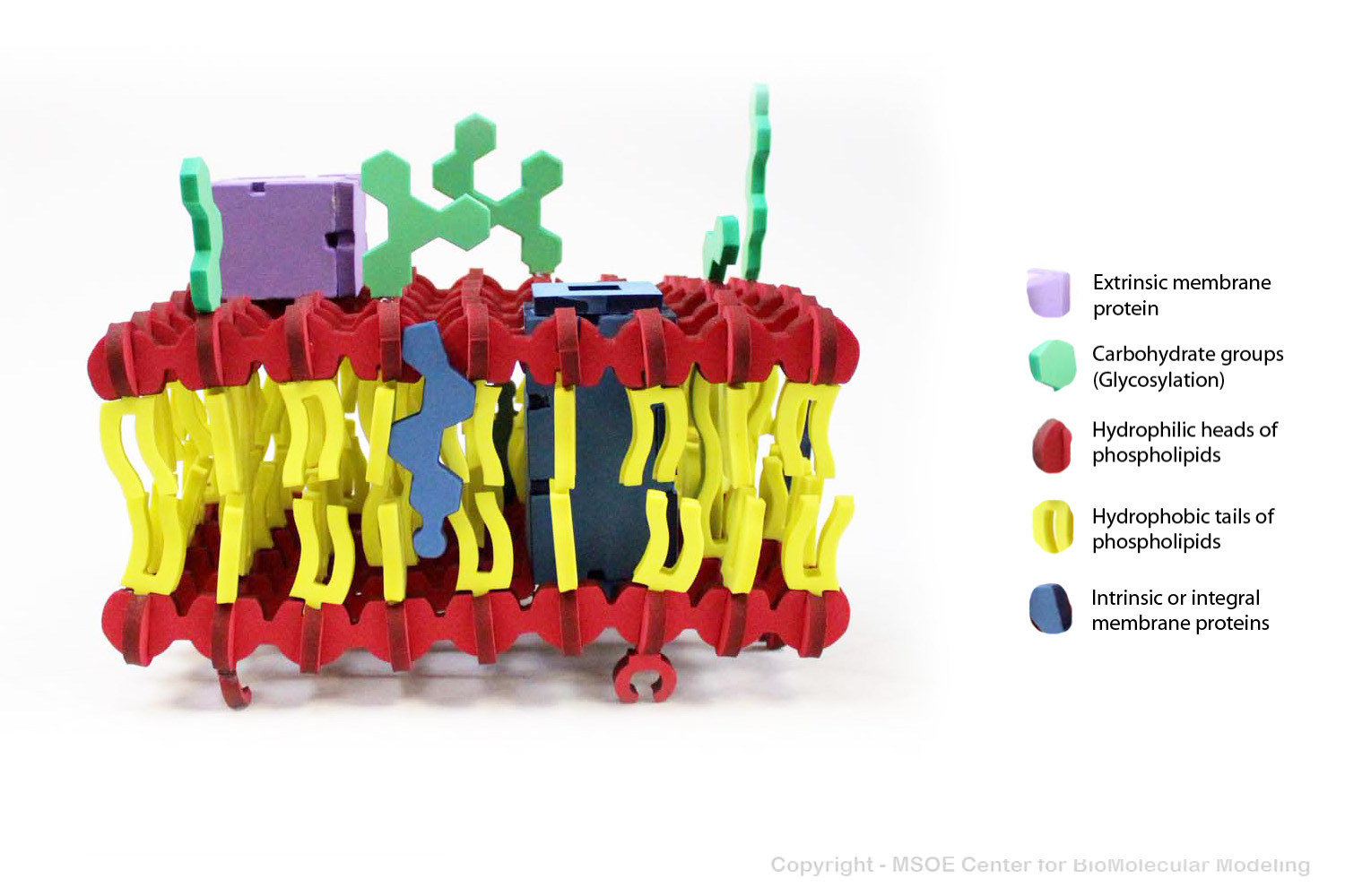
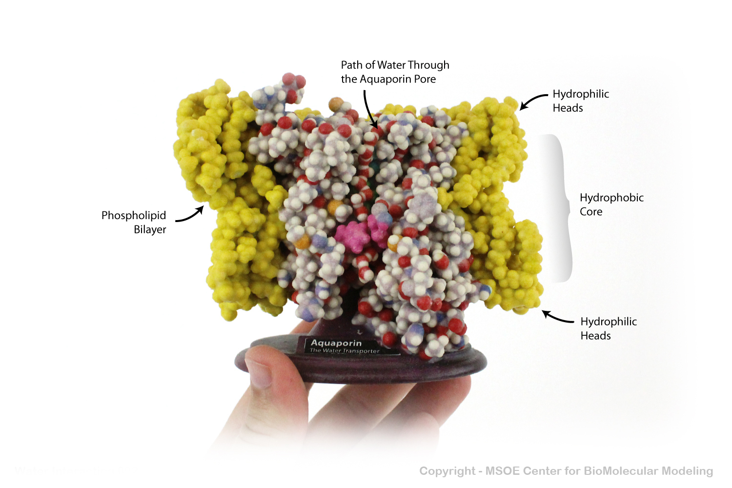
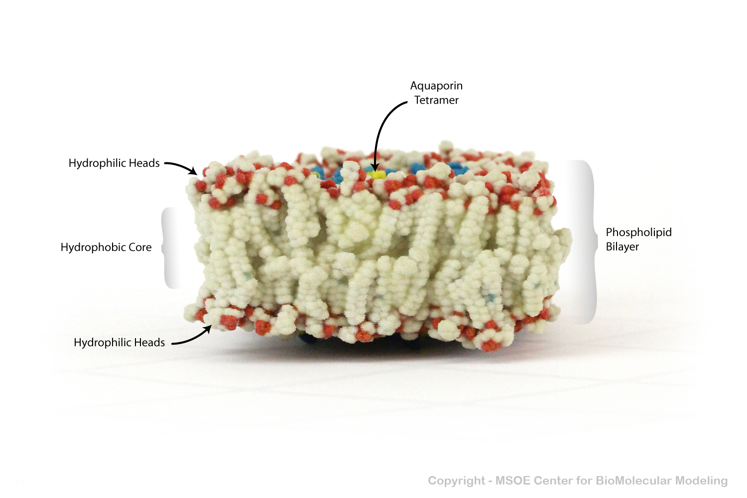
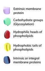
Multiple phospholipids can aggregate into a lipid bilayer, with hydrophobic tails in the middle of the bilayer and hydrophilic heads on the outside of the bilayer. Other molecules, such as protein and glycans decorate the bilayer.