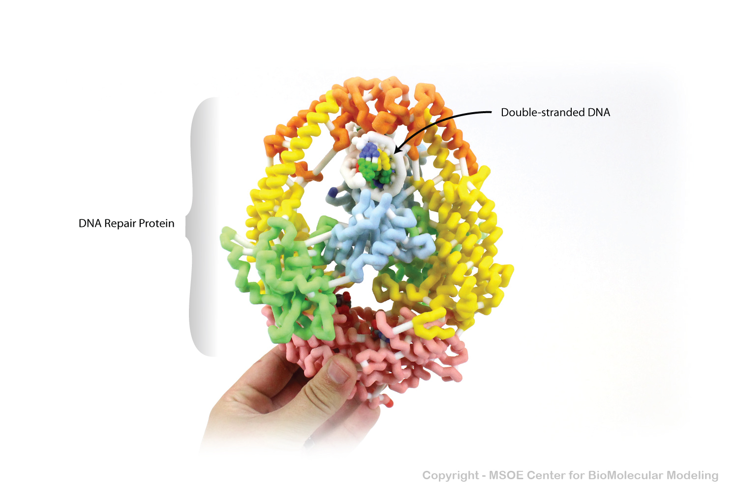
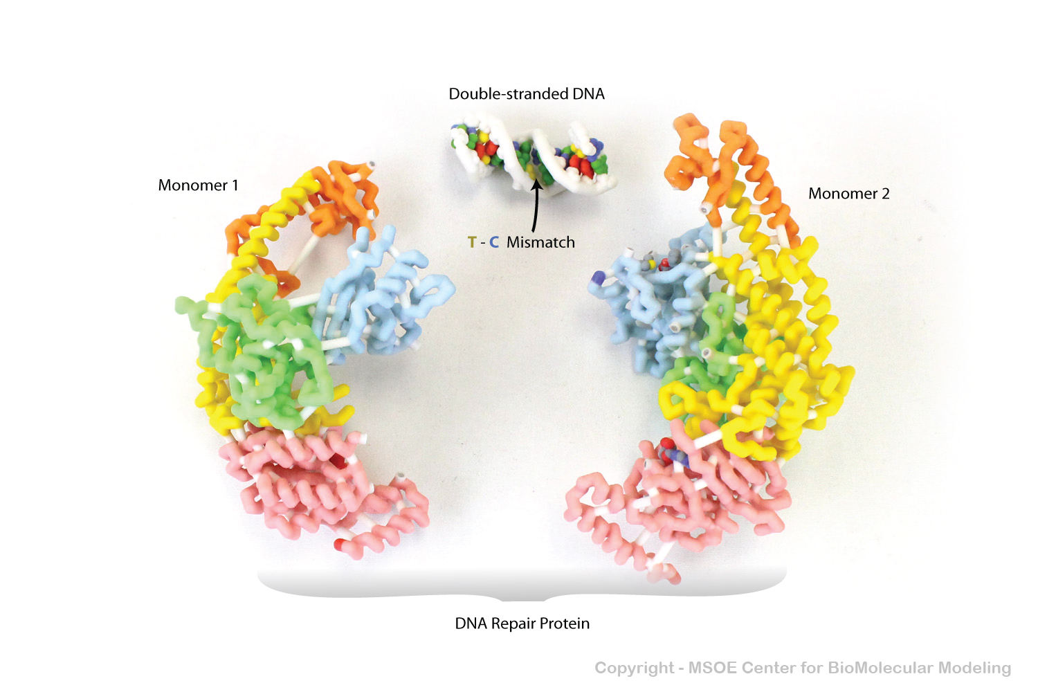

DNA Missmatch Repair Proteins slide along double-stranded DNA looking for "kinks" in the relatively straight DNA molecule. The "kinks" are often the result of missmatched basepairs.
DNA Missmatch Repair Proteins slide along double-stranded DNA looking for "kinks" in the relatively straight DNA molecule. The "kinks" are often the result of missmatched basepairs.
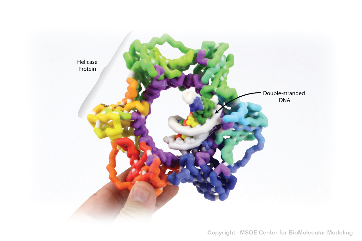
Helicase splits double-stranded DNA into two individual DNA strands during replication. Once split, one strand moves through the hole of the donut-shaped Helicase and the other strand passes outside the hole.
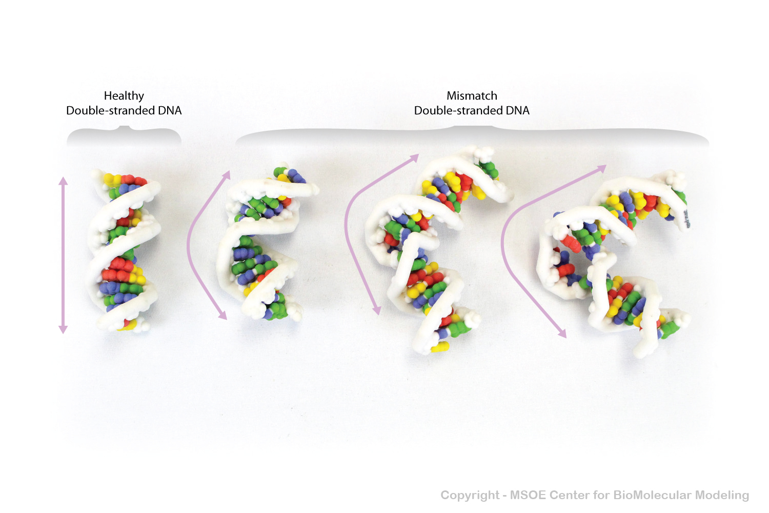
Healthy double-stranded DNA is relatively straight and consistent, while DNA segments with errors or mismatches tend to have kinks or bends, making them irregular and inconsistent.
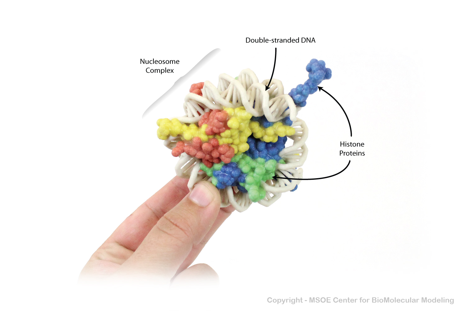
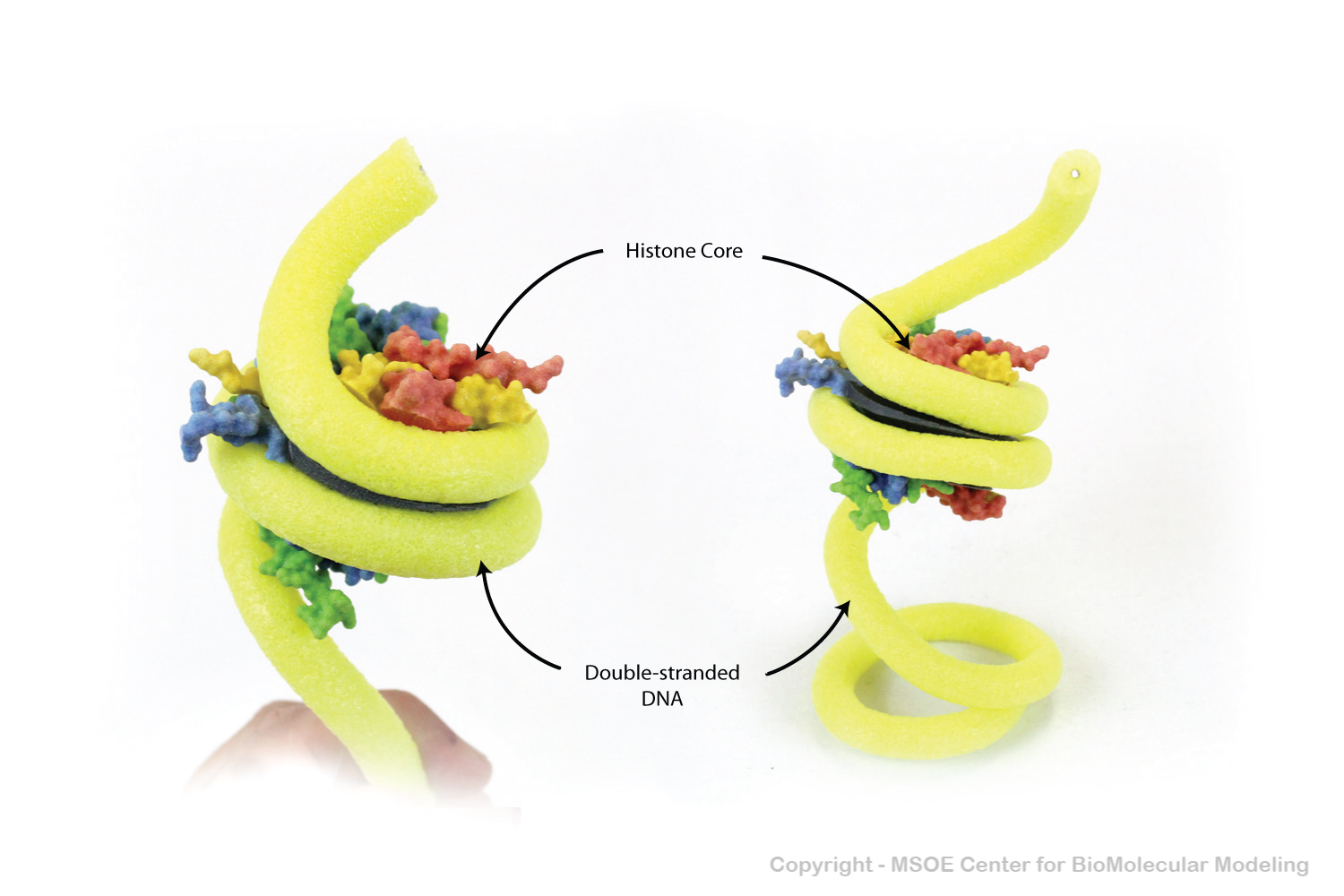
Nucleosomes store and organize double-stranded DNA in the nucleus of all cells. The protein core holds two left-handed wraps of DNA.
Nucleosomes store and organize double-stranded DNA in the nucleus of all cells. The protein core holds two left-handed wraps of DNA.
Nucleosomes store and organize double-stranded DNA in the nucleus of all cells. The protein core holds two left-handed wraps of DNA.
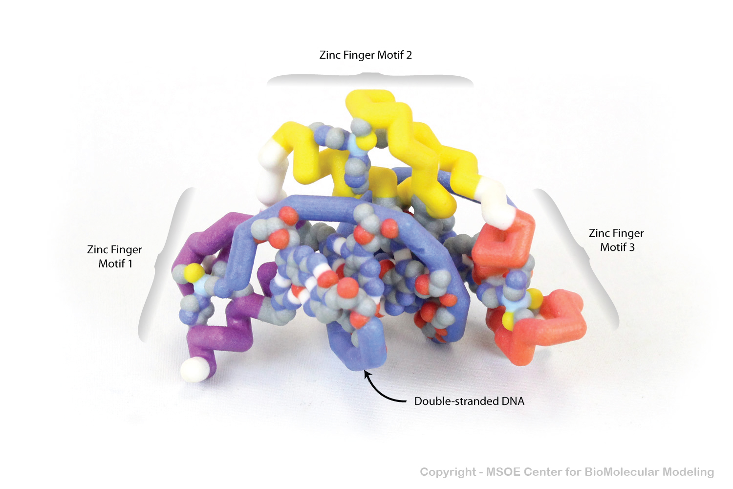
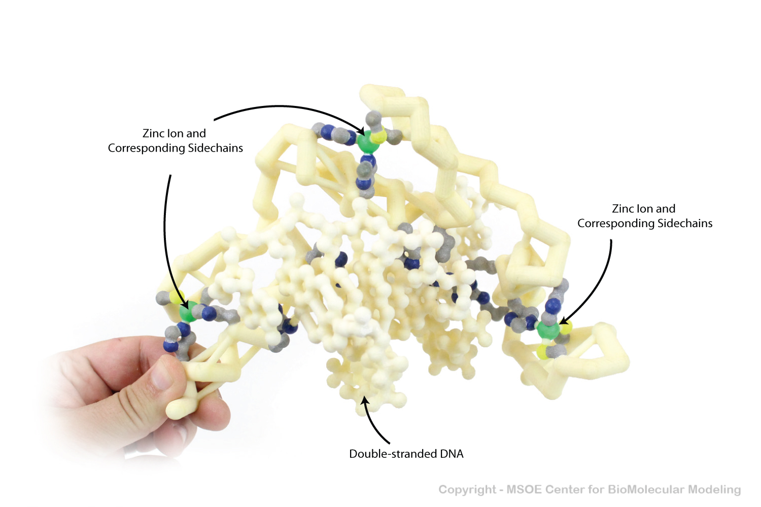
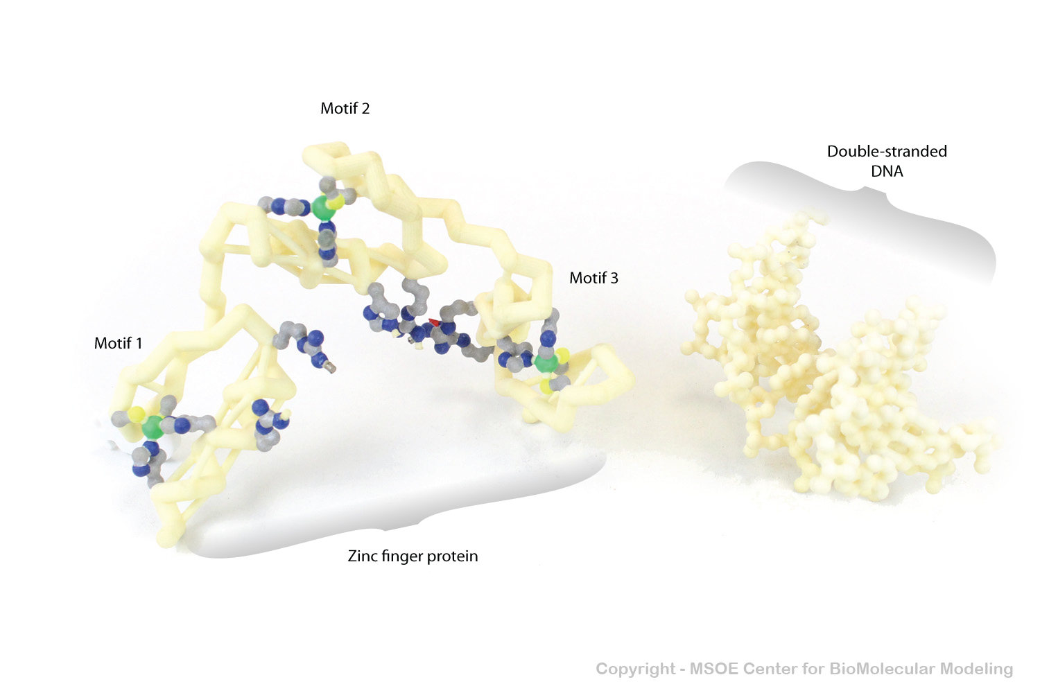
Zinc Finger Motifs are DNA-binding protein folds comprised of one alpha helix and a short two-stranded beta pleated sheet. Multiple Zinc Finger Motifs (in this case, three) often fold together.
Zinc Finger Motifs are DNA-binding protein folds comprised of one alpha helix and a short two-stranded beta pleated sheet. Multiple Zinc Finger Motifs (in this case, three) often fold together.
Zinc Finger Motifs are DNA-binding protein folds comprised of one alpha helix and a short two-stranded beta pleated sheet. Multiple Zinc Finger Motifs (in this case, three) often fold together.
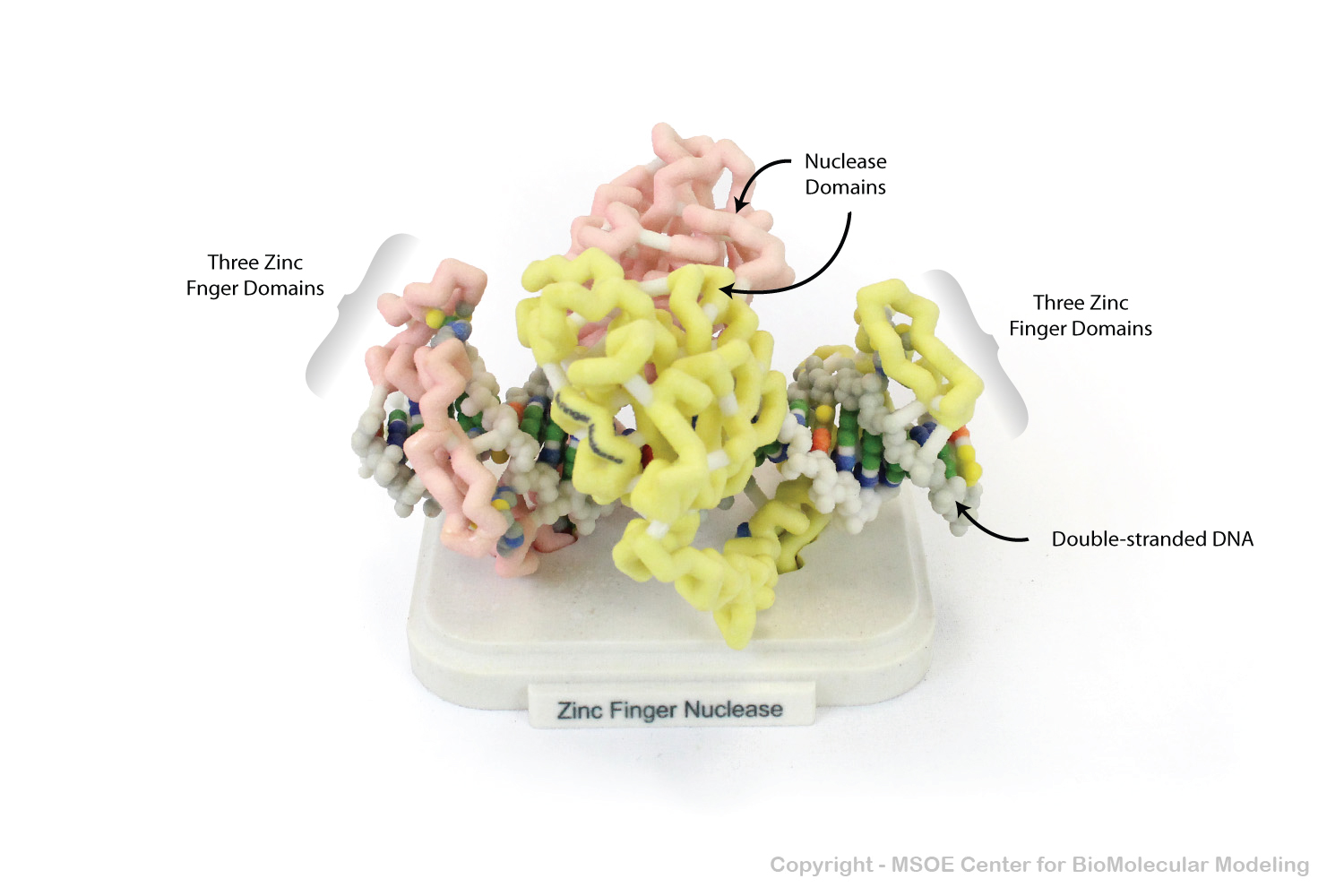
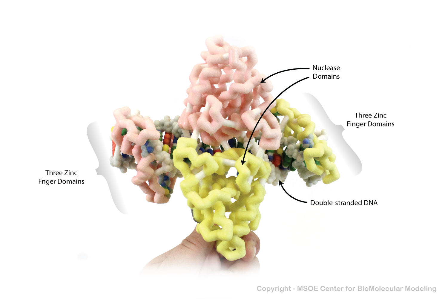
Zinc Finger Nuclease proteins are engineered structures that link zinc finger motifs to nuclease domains. They can bind to a specific sequence of DNA, and then cut the DNA.
Zinc Finger Nuclease proteins are engineered structures that link zinc finger motifs to nuclease domains. They can bind to a specific sequence of DNA, and then cut the DNA.
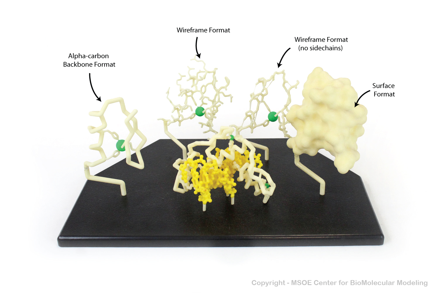
Zinc Finger Motifs are DNA-binding protein folds comprised of one alpha helix and a short two-stranded beta pleated sheet. This model collection shows these motifs in a variety of formats.
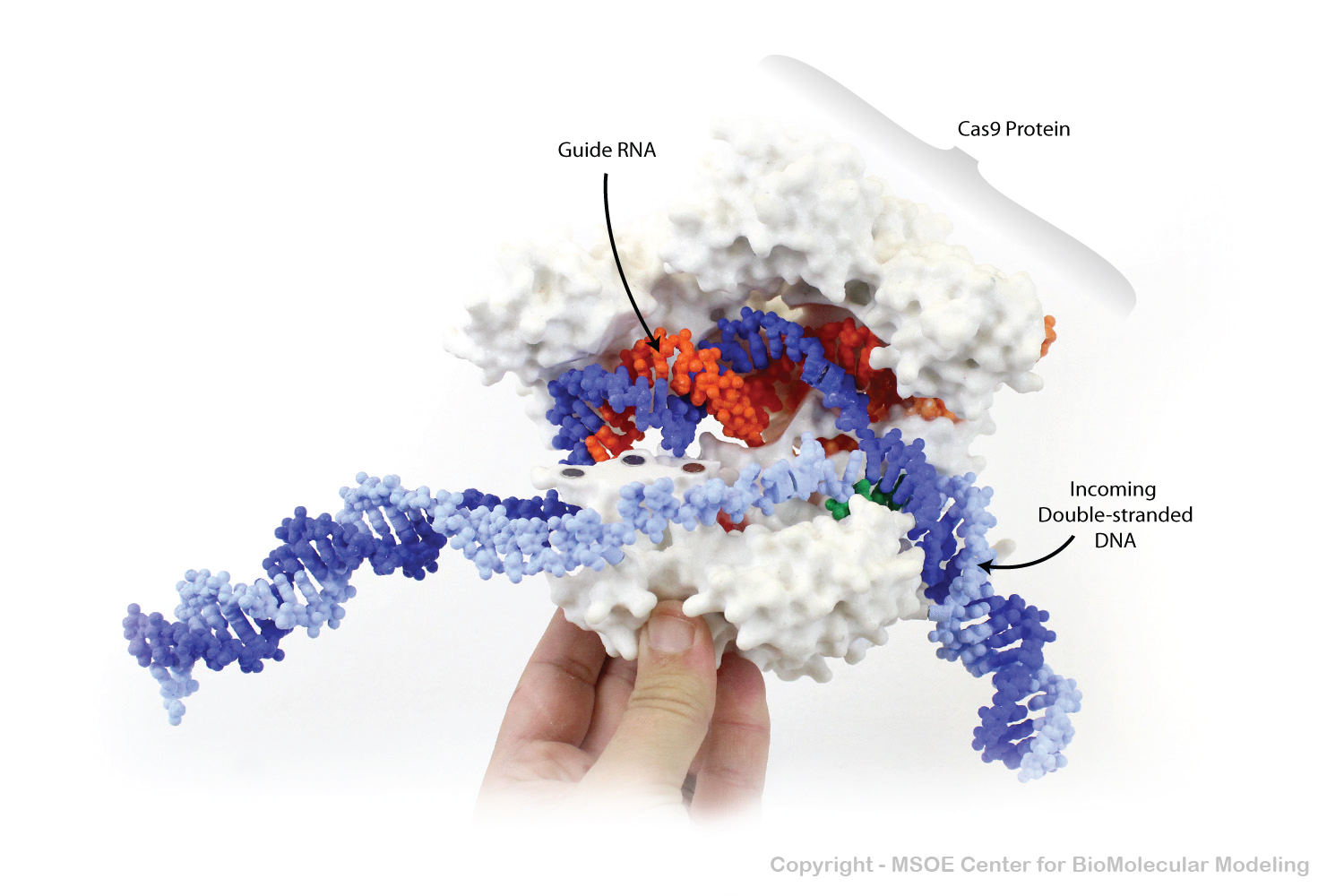
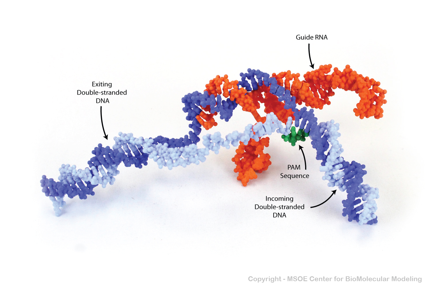
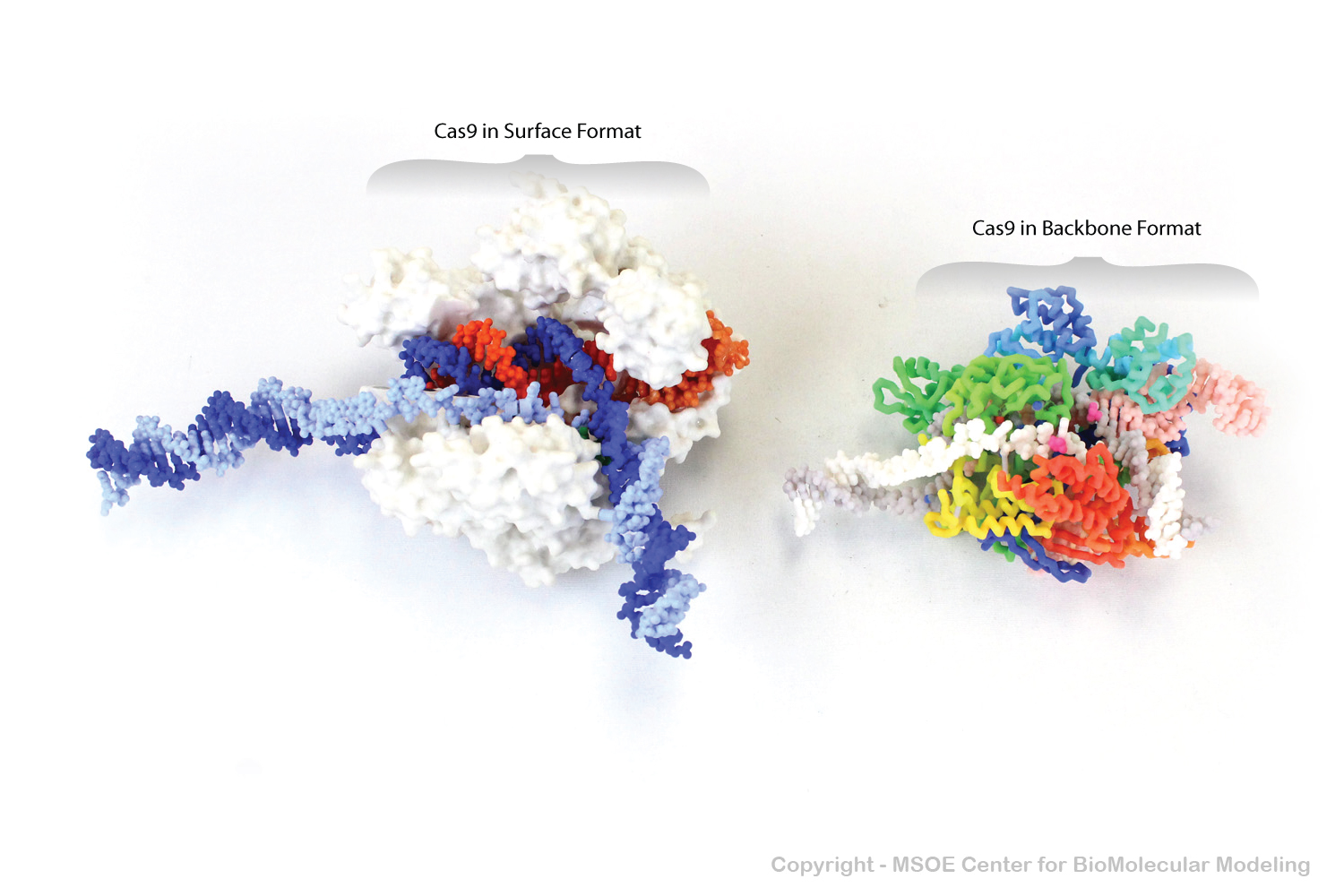
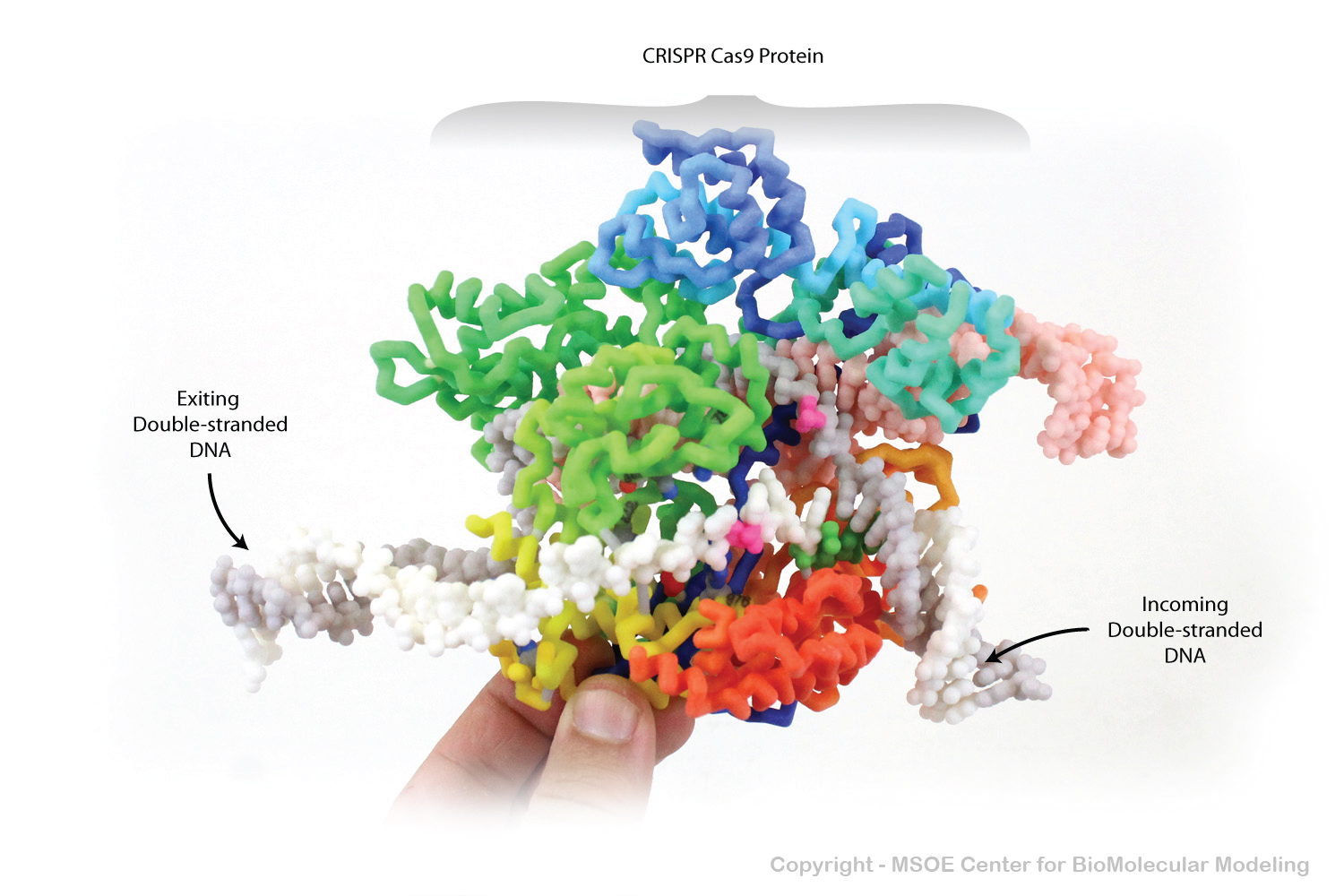
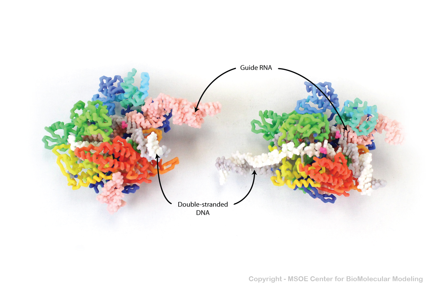
CRSIPR Cas9 is a nuclease protein that is part of the bacterial immune systems. The guide RNA held by the Cas9 protein allows it to find and cut double-stranded DNA at a sequence-specific location.
CRSIPR Cas9 is a nuclease protein that is part of the bacterial immune systems. The guide RNA held by the Cas9 protein allows it to find and cut double-stranded DNA at a sequence-specific location.
CRSIPR Cas9 is a nuclease protein that is part of the bacterial immune systems. The guide RNA held by the Cas9 protein allows it to find and cut double-stranded DNA at a sequence-specific location.
CRSIPR Cas9 is a nuclease protein that is part of the bacterial immune systems. The guide RNA held by the Cas9 protein allows it to find and cut double-stranded DNA at a sequence-specific location.
CRSIPR Cas9 is a nuclease protein that is part of the bacterial immune systems. The guide RNA held by the Cas9 protein allows it to find and cut double-stranded DNA at a sequence-specific location.
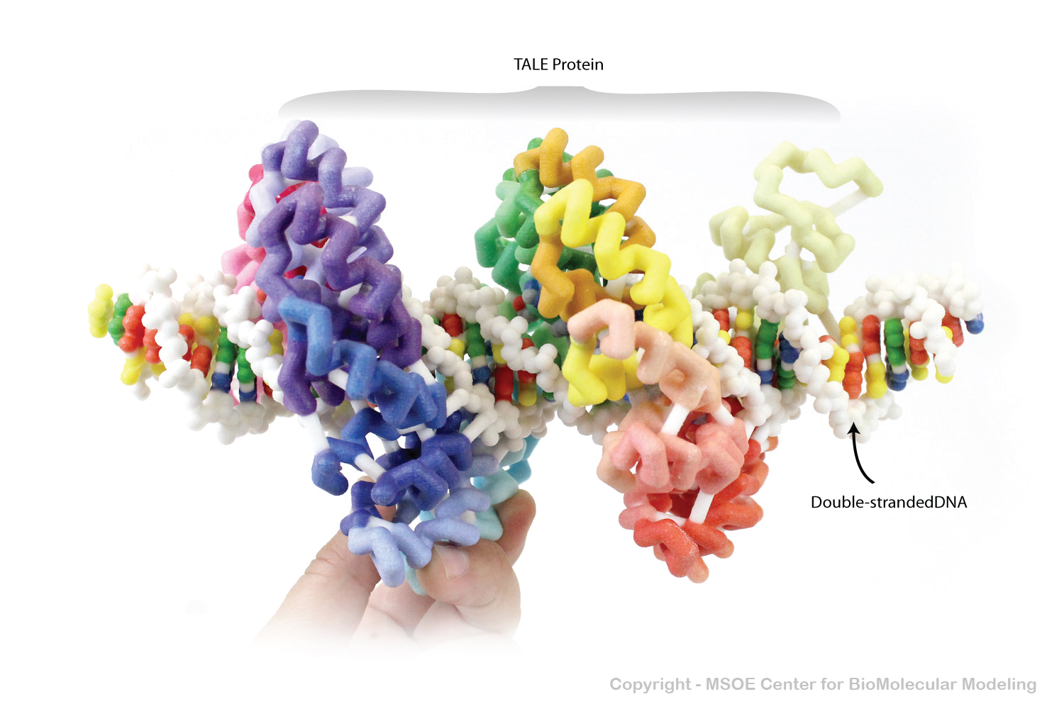
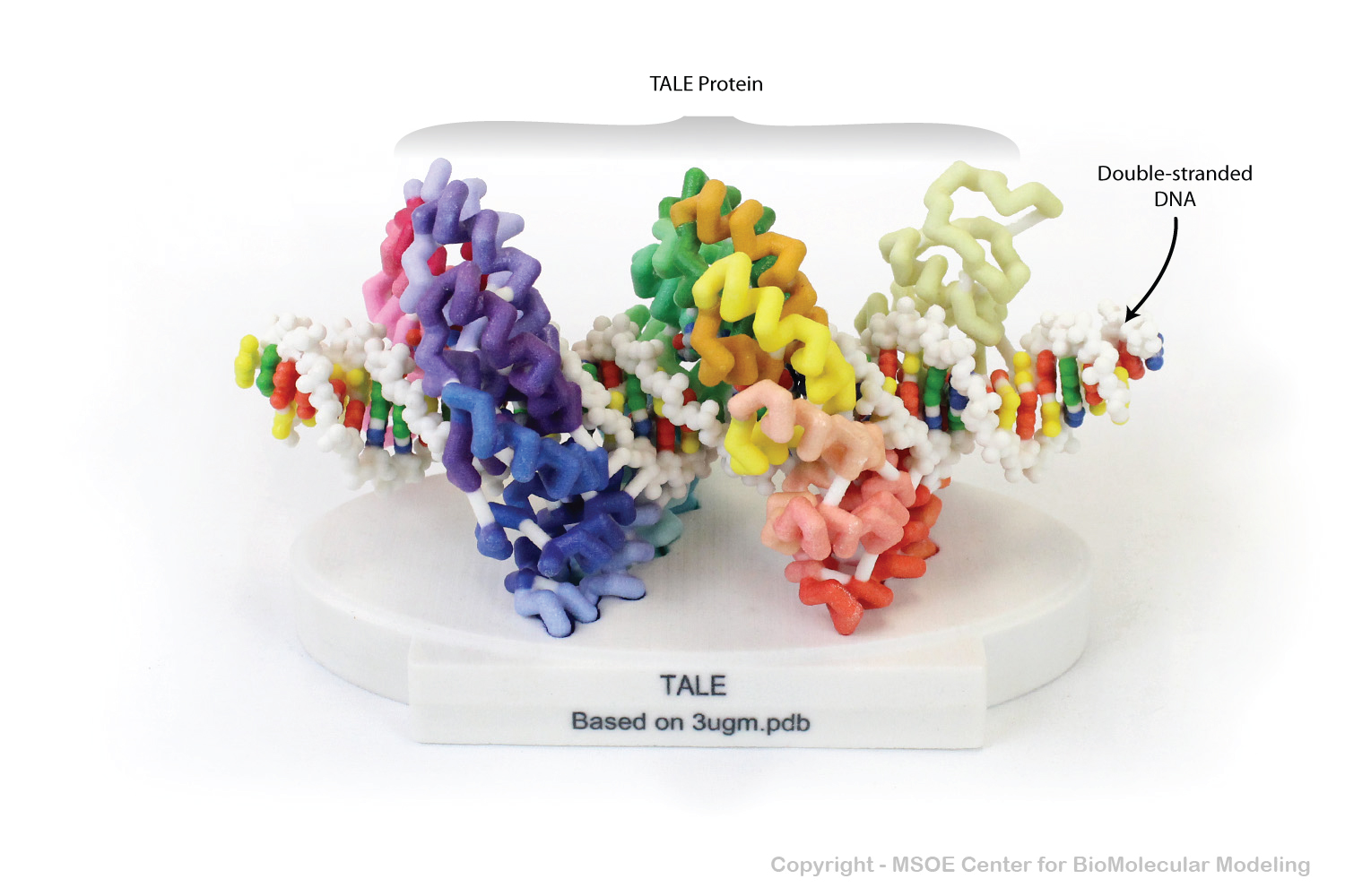
TALE Nuclease proteins are TALE DNA-binding motifs that have been tethered to nuclease protein domains. They can bind to a specific sequence of DNA, and then cut the DNA.
TALE Nuclease proteins are TALE DNA-binding motifs that have been tethered to nuclease protein domains. They can bind to a specific sequence of DNA, and then cut the DNA.
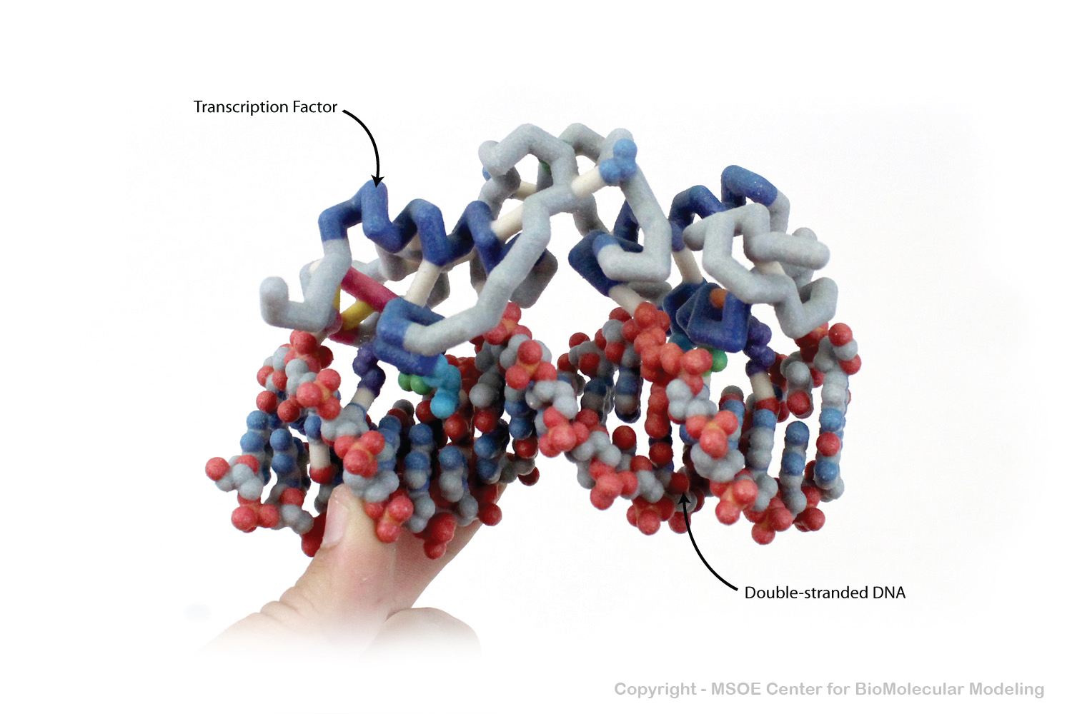
Lac Repressor is a DNA-binding protein that inhibits the expression of genese involved in lactose metabolism. The two chains of the protein both bind in the major groove of double-stranded DNA.
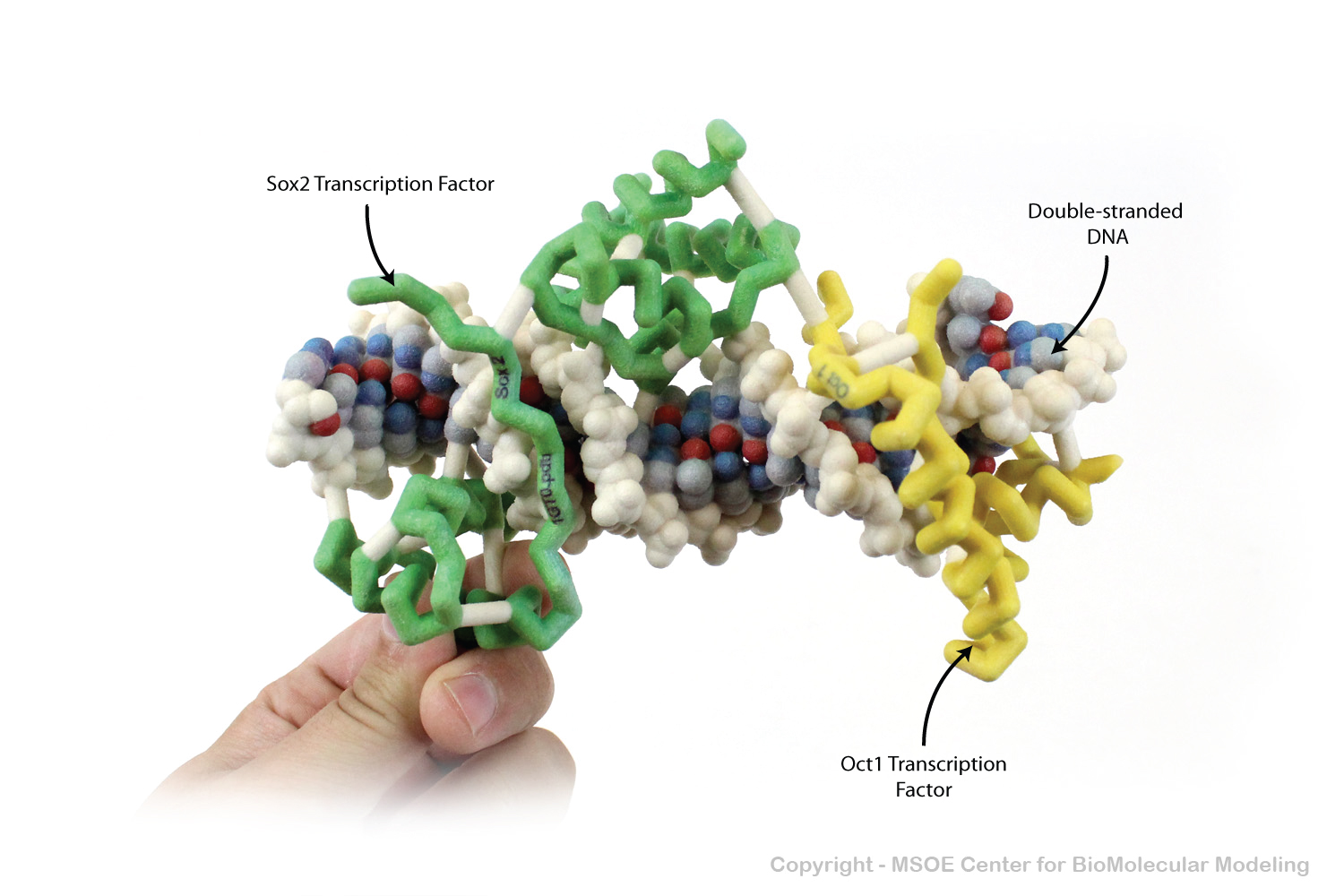
Oct1 and Sox2 are DNA-binding proteins that regulate the expression of genes related to cell groth and apoptosis.
Sox2 is a DNA-binding protein that regulate the expression of genes related to cell groth and apoptosis.
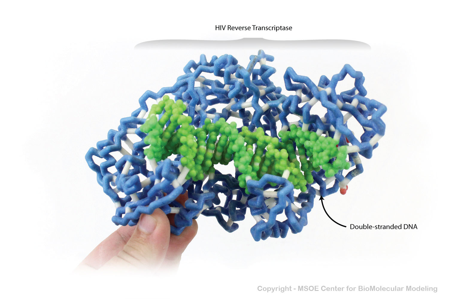
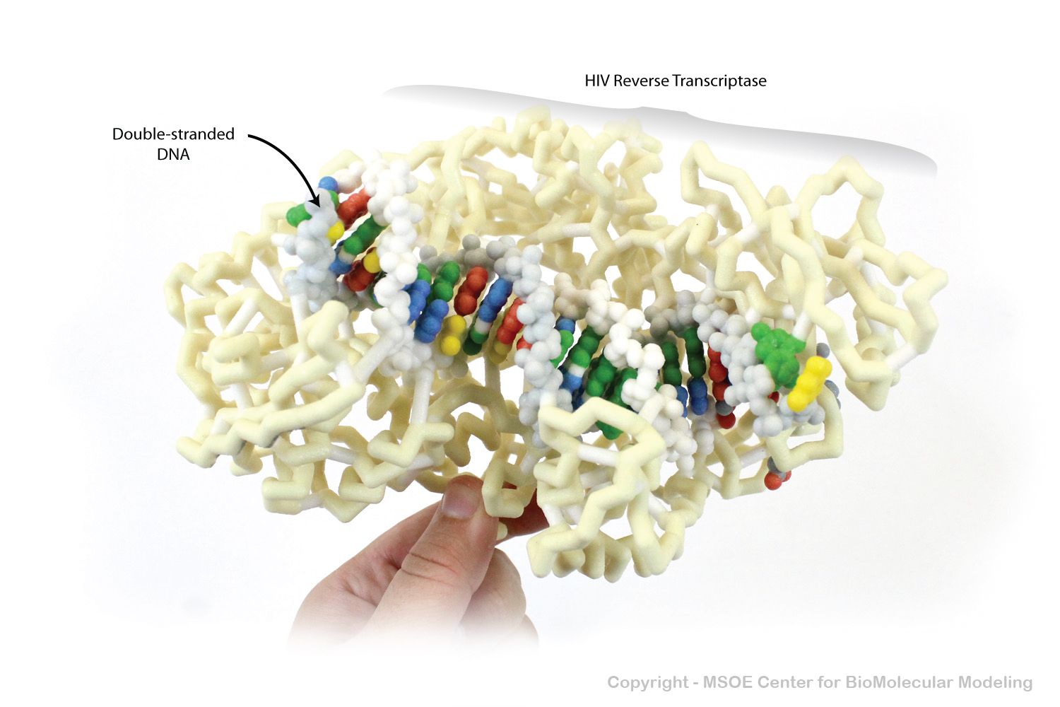
HIV Reverse Transcriptase is a DNA binding protein that can transcribe a single strand of RNA into double-stranded DNA. HIV viruses use this protein to convert their RNA genome into DNA to be used by the host cells to produce more viral proteins.
HIV Reverse Transcriptase is a DNA binding protein that can transcribe a single strand of RNA into double-stranded DNA. HIV viruses use this protein to convert their RNA genome into DNA to be used by the host cells to produce more viral proteins.