

The primary structure of a protein is the specific order of amino acids linked together to form a polypeptide chain.
But polypeptides do not stay straight, as a long chain. Rather, they fold to create complex 3-dimensional shapes.
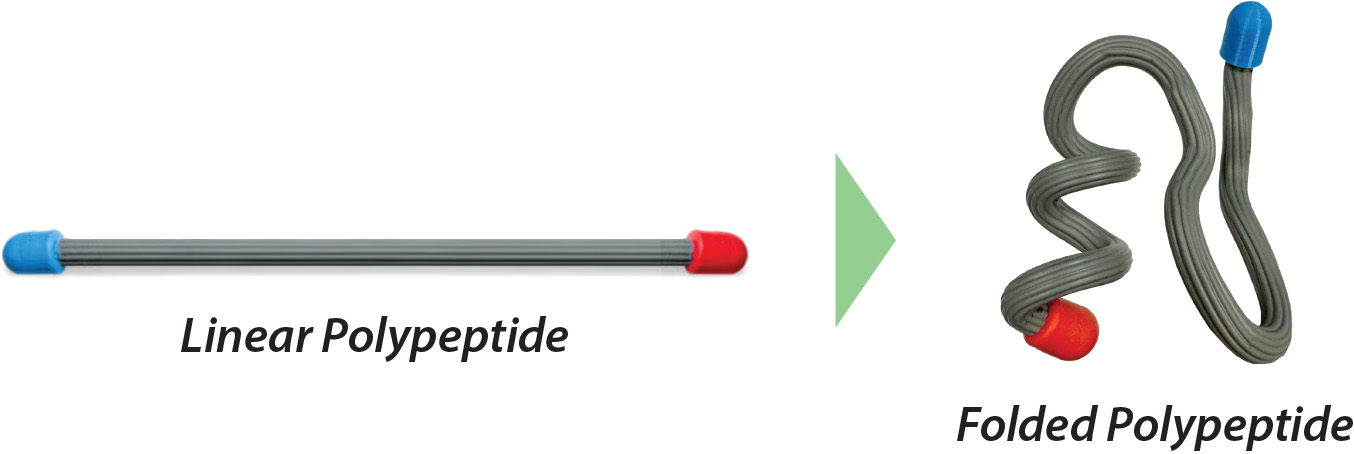

When examining different proteins, you may have noticed that there are two recognizable shapes that are often found throughout a protein's 3-dimensional structure.
One of these shapes looks like a spiral and is called an alpha helix, and the other shape looks like a zig-zag and is called a beta sheet. These two common shapes are referred to as the protein's Secondary Structure.
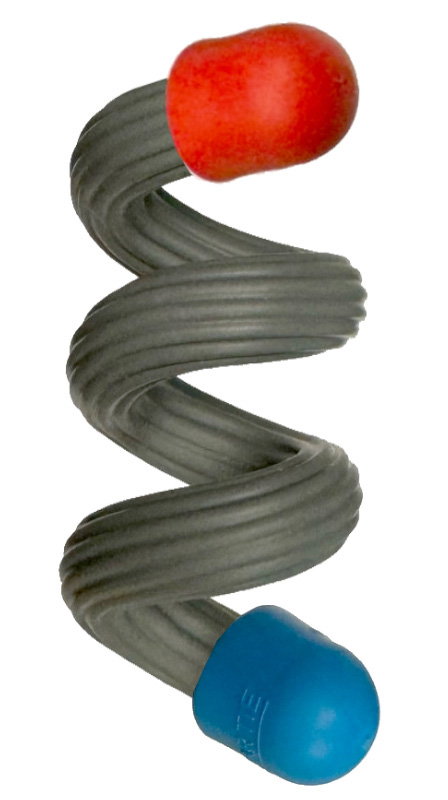
Alpha helices look like a spiral staircase or a spring that coils in a right-handed direction. Show an alpha helix in the interactive display to the right. |
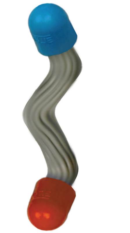
Beta sheets look like a zig-zag structure. Show a beta sheet in the interactive display to the right. |

Alpha helices and beta sheets are supported and reinforced by hydrogen bonds, which often form between the nitrogen atom of one amino acid's backbone and the oxygen atom of another amino acid's backbone.
Click on the links below to see hydrogen bonds represented as yellow cylinders in the two types of secondary structures.
| An alpha helix shown in ball & stick format | A beta sheet shown in ball & stick format |
| An alpha helix shown in backbone format | A beta sheet shown in backbone format |

An example protein called a zinc finger clearly shows both an alpha helix and a beta sheet. Zinc finger proteins help regulate DNA expression in a cell's nucleus. This relatively small protein motif is only 25 amino acids long but includes a four-turn alpha helix and a two strand beta pleated sheet.
Show a zinc finger protein in the interactive display to the right.
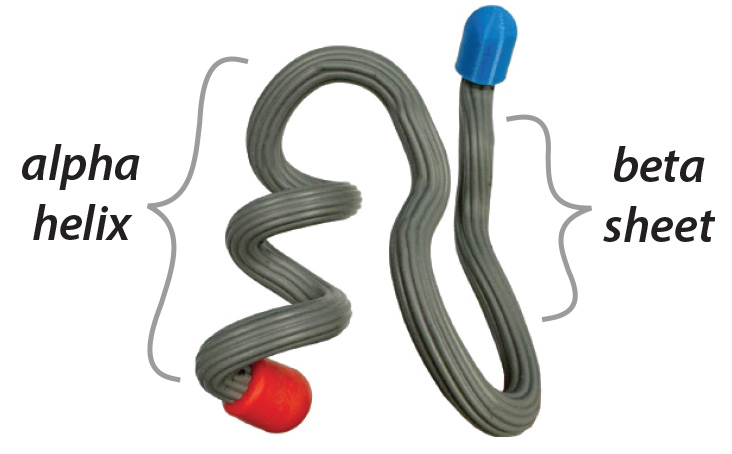

Protein structures can become visually overwhelming. This is especially true for large proteins, which can be thousands of amino acids long and include tens of thousands of atoms!
To avoid confusion proteins images are often shown using different display formats and color schemes. Click on the buttons below to see the zinc finger protein in the interactive display to the right shown in some of the most common display formats and color schemes.
|
Ball & Stick Format displays every single atom in a protein with a small sphere and represents the bonds between these atoms with thin sticks. In Ball & stick format
|
CPK Color Scheme colors every atom by its element type. Carbon atoms are gray, nitrogen atoms are blue, oxygen atoms are red and hydrogen atoms are white.
|
Each of the different display formats and color schemes used in protein visualization has advantages and disadvantages.
For example, spacefill format is an excellent way to represent the overall globular shape of a protein, but may make it difficult to see details at the very center of the protein. Cartoon format is an excellent way to see the overall path of a protein's backbone but it may not show the details of each individual amino acid's R-group.

Click on the images below to view each of the three proteins discussed earlier in the interactive display to the right. Each protein will be colored with the structure color scheme (alpha helices colored magenta and beta sheets colored yellow) to emphasize each protein's unique secondary structures.
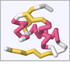 Insulin Proteins help regulate sugar in the bloodstream. |
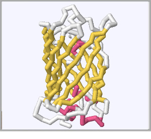 Green Fluorescent Proteins create bioluminescence in animals like jellyfish. |
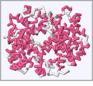 Hemoglobin Proteins safely carry oxygen in the blood. |


© Copyright 1995- - 3D Molecular Designs