Click Above to Play the Video
Proteins are large, complex molecules that are made in the polar, watery environment of the cell. They are made by joining amino acids together in a particular sequence, referred to as the protein’s Primary Structure.
Because each of the 20 types of amino acids is different in shape and chemical property, proteins fold up into amazingly complex 3-dimensional shapes based on their specific amino acid sequence, and following basic principles of chemistry.
Click to See a Chart of these 20 Types of Amino Acids ▶

We are going to examine these basic principles of chemistry using betaglobin, a 146 amino acid long protein, shown in the interactive Jmol window to the right
The betaglobin protein is currently displayed in "spacefilled format", in which each atom is shown as a color-coded sphere:
- Carbon atoms are grey
- Oxygen atoms are red
- Nitrogen atoms are blue
- Sulfur atoms are yellow
This "spacefill format" shows the overall globular shape of the betaglobin protein, but is not very useful in seeing how the linear sequence of 146 amino acids folds up into a compact shape.
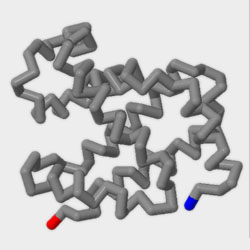
But if we fade out all of the atoms except the alpha carbon of each amino acid. . . and then connect each alpha carbon with a cylinder. . . we see a much less cluttered representation of the protein, including a blue cap for the beginning or N-terminus of the protein chain - and a red cap for the end or C-terminus.
In this “backbone format” we can also see that the protein has folded into a series of alpha helices (the protein’s secondary structure) – which in turn fold up into a compact, globular shape (the protein’s tertiary structure).
For now, we are going to return to "spacefill format" and ignore the alpha helix secondary structures that are more easily visible in “backbone format”. Instead, we will focus on the tertiary structure – the overall globular shape of the betaglobin protein
Hydrophobic and Hydrophilic
The First Principle of Chemistry of Drives Protein Folding
Click Above to Play the Video
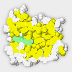
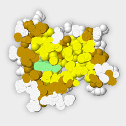
If we color all of the hydrophobic amino acids yellow, and all of the hydrophilic amino acids white, we can begin to explore their location and distribution throughout the betaglobin protein.
- Hydrophobic amino acids are yellow
- Hydrophilic amino acids are white
The first principle of chemistry is that hydrophobic (water-fearing) amino acid sidechains tend to fold to the inside of a globular protein, where their interaction with hydrophilic water is minimized.
At the same time, hydrophilic (water-loving) amino acid sidechains tend to fold to the outside of the globular protein, where they can freely interact with water in the aqueous environment of the cell’s cytoplasm.
This view does not allow us to see what is buried deep inside the protein. . . but by “slabbing” the previous view, we have cut through the protein with a plane that is parallel with the screen, and removed that part of the protein that is in front of the slab plane.
Note also that you can rotate the protein through this slab plane by left-clicking and dragging in the Jmol window. And you can use the slider bar below to change the “depth” of the slab plane across the protein.
This view convincingly demonstrates that the core of this protein is tightly packed with hydrophobic amino acids, and most of the exterior surface of the protein is covered with hydrophilic amino acids.
If you are skeptical (like all scientists), you may have noticed that there are places on the exterior surface of the protein where some hydrophobic amino acids are exposed to the hydrophilic, watery environment of the cell.
- Hydrophobic amino acids in the protein's core are yellow
- Hydrophobic amino acids on the protein's exterior are dark yellow
- Hydrophilic amino acids are white
Betaglobin appears to be a lot like a teenager – it doesn’t follow all the rules all of the time. In biology, when you encounter an exception to a generally followed rule, you should get excited. There is probably a reason for the exception, and if you can understand why the rule was not followed, you will learn something.
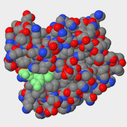
If we rotate the betaglobin structure to find the area with the greatest concentration of yellow hydrophobic amino acids exposed on the surface. You will see one especially large exposed hydrophobic sidechain.
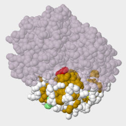
Perhaps there is more to this protein than meets the eye. . . That’s right, the betaglobin protein does not exist as a single protein chain, but as part of a four chain complex known as hemoglobin. When a protein is comprised of more than one amino acid chain, it is said to have quaternary structure.
And here is the important part. . . when two protein chains interact to form quaternary structure, these interacting surfaces are usually hydrophobic in nature. . . And you have just learned something!
Charged Residues
The Second Principle of Chemistry of Drives Protein Folding
Click Above to Play the Video
A second basic principle of chemistry that drives protein folding is that positively-charged amino acid sidechains want to be close to negatively-charged sidechains, forming what is called a “Salt Bridge”.
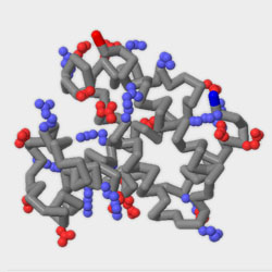
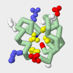
To help us visualize potential salt bridges in the betaglobin protein, lets return to “backbone format”, in which only the alpha carbon atoms for each amino acid are shown – connected by cylinders. Then, we can add the positively charged sidechains in blue, and the negatively charged sidechains in red.
These electrostatic interactions between oppositely-charged sidechains are often referred to as “salt bridges”.
Only 38% of the charged sidechains are involved in salt bridges. As a result, we can conclude that while this principle of chemistry is used to stabilize the folded structure of a protein, only a subset of the charged amino acids are involved in these interactions.
Conclusion
To the Principles of Chemistry of Drive Protein Folding
Click Above to Play the Video