
Basic Principles of Chemistry that Drive Protein Folding - part 1
Basic Principles of Chemistry that Drive Protein Folding - part 1


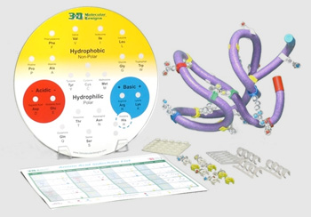
Proteins are large molecules that are synthesized in the polar, watery environment of the cell. They are made by joining amino acids together in a particular sequence. Because each of the 20 amino acids is different in shape and chemical property, proteins fold up into different 3-dimensional shapes following basic principles of chemistry.
Click on each of the proteins shown below to view them in the Jmol molecular display window to the right.
Note that the Jmol display to the right is fully interactive and can be rotated by clicking and dragging with your mouse!
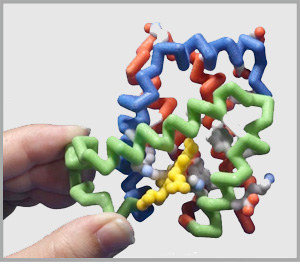 ß-globin Proteins safely carry oxygen in the blood. |
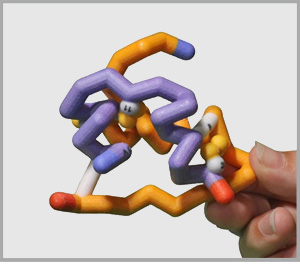 Insulin Proteins help regulate sugar in the bloodstream. |
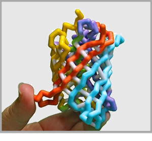 Green Fluorescent Proteins create bioluminescence in animals like jellyfish. |

Click Here to load the Betaglobin Protein
The Amino Acid Starter Kit from 3D Molecular Designs introduces the basic principles of chemistry that drive protein folding. This Jmol tutorial will allow you to determine how accurately a real protein, ß-globin, reflects these concepts in its final, folded structure.
The ß-globin protein shown to the right is in spacefill format, in which each atom is represented by a sphere the diameter of the atom's electron cloud. The protein has been colored with the CPK color scheme, meaning each type of element will have a unique color assinged to it.
The first prinicple of chemistry that drives protein folding suggests that hydrophobic amino acids should be buried inside the protein where they can hide away from the water that surrounds the protein.
Simultaneously, hydrophillic (polar and charged) amino acids should be on the surface of the folded protein where they are exposed to and can interact with water.
|
Hydrophobic |
Hydrophillic |
To make it easier to visualize where the hydrophobic and hydrophillic amino acids are on the ß-globin protein, click the buttons below to apply a more visually useful color scheme.
Click Here to color the Hydrophobic Amino Acids Yellow and Hydrophillic Amino Acids White
The CPK colored molecule that is buried in the ß-globin is known as the heme group. The orange colored atom in its center is iron (Fe) and binds to oxygen gas (O2). What is the function of the ß-globin protein?
Notice that proteins are a lot like students (and most adults!) in that they don't follow all of the rules all of the time. Can you see examples of hydrophobic amino acids that are exposed on the surface of ß-globin?
To get a better look at the inside of the protein, use the button below to slab the protein. Slabbing a molecule in a 3-dimensional Jmol display visually cuts it in half, making the half of it visually closest to invisible
Click Here to slab the ß-globin protein
Note that you can change the slab depth by holding the Ctrl and Shift keys and drag up and down using the left mouse button.
Answer the question below based on your analysis of the ß-globin protein. Clicking on the question will display the correct answer.
Pretty well. Although there are a few hydrophobic amino acids that are exposed on the surface, the central core of the protein, seen in the slabbed view, is entirely hydrophobic.
The second principle of chemistry that drives protein folding suggests that charged amino acids will be on the surface of a globular protein and that positively-charged amino acidss will often be paired with negatively-charged amino acids.
Positive and Negative
Yes, all of the charged amino acids - both positively-charged and negatively-charged - are exposed on the outter surface of the ß-globin protein.
Rotate this final image to examine closely how the positively-charged and negatively-charged amino acids are positioned in this protein.
Sometimes they are paired, sometimes they are not. There are three pairs of positively-charged and negatively-charged amino acids on the surface of this protein. The rest of the charged amino acids do not make pairs.
© Copyright 1995- - MSOE Center for BioMolecular Modeling